Glial fibrillary acidic protein,GFAP
对比
货号-规格 货期 价格 数量
abs131715-10uL 1-2周
260 .00
abs131715-25uL 1-2周
600 .00
abs131715-100uL 1-2周
2100 .00
abs131715-500uL 1-2周
5280 .00
abs131715-1mL 1-2周
询价
订购信息:
您可以从我们的授权经销商处购买absin产品或获得技术支持。若要查看您所在地区的经销商,请从以下的下拉列表中选择。
实验结果图
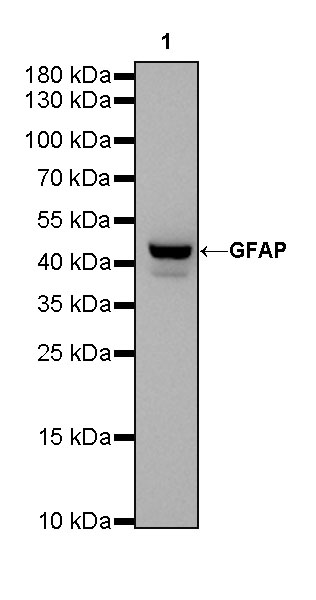
WB result of GFAP Rabbit mAb Primary antibody: GFAP Rabbit mAb at 1/1000 dilution Lane 1: mouse brain lysate 20 µg Secondary antibody: Goat Anti-Rabbit IgG, (H+L), HRP conjugated at 1/10000 dilution Predicted MW: 50kDa Observed MW: 42kDa Exposure time: 30s
WB result of GFAP Rabbit mAb Primary antibody: GFAP Rabbit mAb at 1/1000 dilution Lane 1: rat brain lysate 20 µg Secondary antibody: Goat Anti-Rabbit IgG, (H+L), HRP conjugated at 1/10000 dilution Predicted MW: 50kDa Observed MW: 50kDa Exposure time: 30s
IHC shows positive staining in paraffin-embedded human cerebellum. Anti-GFAP antibody was used at 1/1000 dilution, followed by a HRP Polymer for Mouse & Rabbit IgG (ready to use). Counterstained with hematoxylin. Heat mediated antigen retrieval with Tris/EDTA buffer pH9.0 was performed before commencing with IHC staining protocol.
IHC shows positive staining in paraffin-embedded rat colon. Anti-GFAP antibody was used at 1/1000 dilution, followed by a HRP Polymer for Mouse & Rabbit IgG (ready to use). Counterstained with hematoxylin. Heat mediated antigen retrieval with Tris/EDTA buffer pH9.0 was performed before commencing with IHC staining protocol.
IHC shows positive staining in paraffin-embedded human cerebral cortex. Anti-GFAP antibody was used at 1/1000 dilution, followed by a HRP Polymer for Mouse & Rabbit IgG (ready to use). Counterstained with hematoxylin. Heat mediated antigen retrieval with Tris/EDTA buffer pH9.0 was performed before commencing with IHC staining protocol.
IHC shows positive staining in paraffin-embedded human glioma. Anti-GFAP antibody was used at 1/1000 dilution, followed by a HRP Polymer for Mouse & Rabbit IgG (ready to use). Counterstained with hematoxylin. Heat mediated antigen retrieval with Tris/EDTA buffer pH9.0 was performed before commencing with IHC staining protocol.
IHC shows positive staining in paraffin-embedded human astrocytoma. Anti-GFAP antibody was used at 1/1000 dilution, followed by a HRP Polymer for Mouse & Rabbit IgG (ready to use). Counterstained with hematoxylin. Heat mediated antigen retrieval with Tris/EDTA buffer pH9.0 was performed before commencing with IHC staining protocol.
IHC shows positive staining in paraffin-embedded human breast cancer. Anti-GFAP antibody was used at 1/1000 dilution, followed by a HRP Polymer for Mouse & Rabbit IgG (ready to use). Counterstained with hematoxylin. Heat mediated antigen retrieval with Tris/EDTA buffer pH9.0 was performed before commencing with IHC staining protocol.
Negative control: IHC shows negative staining in paraffin-embedded human skeletal muscle. Anti-GFAP antibody was used at 1/1000 dilution, followed by a HRP Polymer for Mouse & Rabbit IgG (ready to use). Counterstained with hematoxylin. Heat mediated antigen retrieval with Tris/EDTA buffer pH9.0 was performed before commencing with IHC staining protocol.
IHC shows positive staining in paraffin-embedded mouse cerebral cortex. Anti-GFAP antibody was used at 1/1000 dilution, followed by a HRP Polymer for Mouse & Rabbit IgG (ready to use). Counterstained with hematoxylin. Heat mediated antigen retrieval with Tris/EDTA buffer pH9.0 was performed before commencing with IHC staining protocol.
IHC shows positive staining in paraffin-embedded mouse stomach. Anti-GFAP antibody was used at 1/1000 dilution, followed by a HRP Polymer for Mouse & Rabbit IgG (ready to use). Counterstained with hematoxylin. Heat mediated antigen retrieval with Tris/EDTA buffer pH9.0 was performed before commencing with IHC staining protocol.
IHC shows positive staining in paraffin-embedded rat cerebral cortex. Anti-GFAP antibody was used at 1/1000 dilution, followed by a HRP Polymer for Mouse & Rabbit IgG (ready to use). Counterstained with hematoxylin. Heat mediated antigen retrieval with Tris/EDTA buffer pH9.0 was performed before commencing with IHC staining protocol.

WB resuLt of GFAP Rabbit mAb 
Primary antibody: GFAP Rabbit mAb at 1/1000 dilution
Lane 1: mouse brain lysate 20 µg
Secondary antibody: Goat Anti-Rabbit IgG, (H+L), HRP conjugated at 1/10000 dilution
Predicted MW: 50kDa
Observed MW: 42kDa


WB resuLt of GFAP Rabbit mAb 
Primary antibody: GFAP Rabbit mAb at 1/1000 dilution
Lane 1: rat brain lysate 20 µg
Secondary antibody: Goat Anti-Rabbit IgG, (H+L), HRP conjugated at 1/10000 dilution
Predicted MW: 50kDa
Observed MW: 50kDa
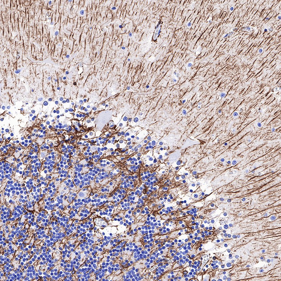
IHC shows positive staining in paraffin-embedded human cerebellum. Anti-GFAP antibody was used at 1/1000 dilution, followed by a HRP Polymer for Mouse & Rabbit IgG (ready to use). Counterstained with hematoxylin. Heat mediated antigen retrieval with Tris/EDTA buffer pH9.0 was performed before commencing with IHC staining protocol.
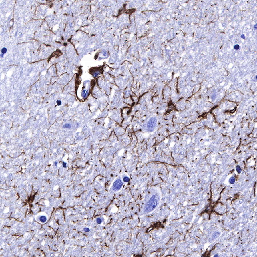
IHC shows positive staining in paraffin-embedded human cerebral cortex. Anti-GFAP antibody was used at 1/1000 dilution, followed by a HRP Polymer for Mouse & Rabbit IgG (ready to use). Counterstained with hematoxylin. Heat mediated antigen retrieval with Tris/EDTA buffer pH9.0 was performed before commencing with IHC staining protocol.
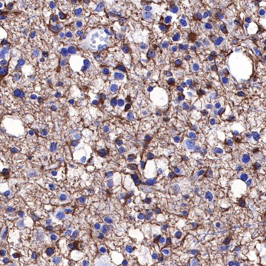
IHC shows positive staining in paraffin-embedded human glioma. Anti-GFAP antibody was used at 1/1000 dilution, followed by a HRP Polymer for Mouse & Rabbit IgG (ready to use). Counterstained with hematoxylin. Heat mediated antigen retrieval with Tris/EDTA buffer pH9.0 was performed before commencing with IHC staining protocol.

IHC shows positive staining in paraffin-embedded human astrocytoma. Anti-GFAP antibody was used at 1/1000 dilution, followed by a HRP Polymer for Mouse & Rabbit IgG (ready to use). Counterstained with hematoxylin. Heat mediated antigen retrieval with Tris/EDTA buffer pH9.0 was performed before commencing with IHC staining protocol.

IHC shows positive staining in paraffin-embedded human breast cancer. Anti-GFAP antibody was used at 1/1000 dilution, followed by a HRP Polymer for Mouse & Rabbit IgG (ready to use). Counterstained with hematoxylin. Heat mediated antigen retrieval with Tris/EDTA buffer pH9.0 was performed before commencing with IHC staining protocol.

Negative control: IHC shows negative staining in paraffin-embedded human skeletal muscle. Anti-GFAP antibody was used at 1/1000 dilution, followed by a HRP Polymer for Mouse & Rabbit IgG (ready to use). Counterstained with hematoxylin. Heat mediated antigen retrieval with Tris/EDTA buffer pH9.0 was performed before commencing with IHC staining protocol.
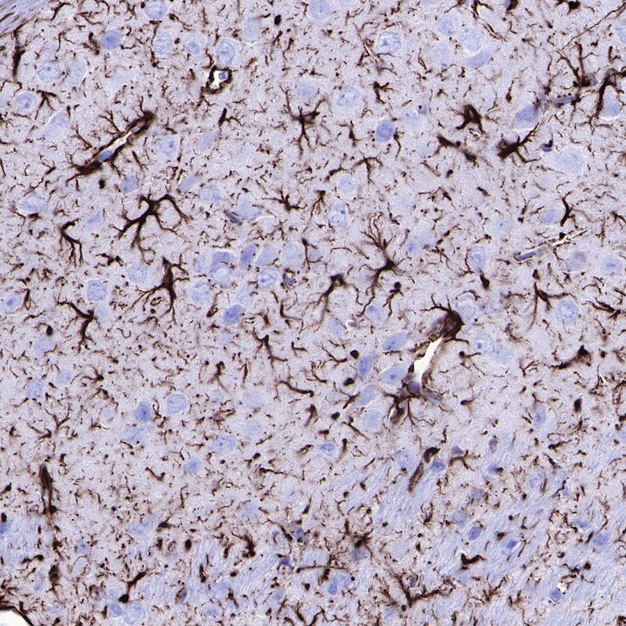
IHC shows positive staining in paraffin-embedded mouse cerebral cortex. Anti-GFAP antibody was used at 1/1000 dilution, followed by a HRP Polymer for Mouse & Rabbit IgG (ready to use). Counterstained with hematoxylin. Heat mediated antigen retrieval with Tris/EDTA buffer pH9.0 was performed before commencing with IHC staining protocol.

IHC shows positive staining in paraffin-embedded mouse stomach. Anti-GFAP antibody was used at 1/1000 dilution, followed by a HRP Polymer for Mouse & Rabbit IgG (ready to use). Counterstained with hematoxylin. Heat mediated antigen retrieval with Tris/EDTA buffer pH9.0 was performed before commencing with IHC staining protocol.
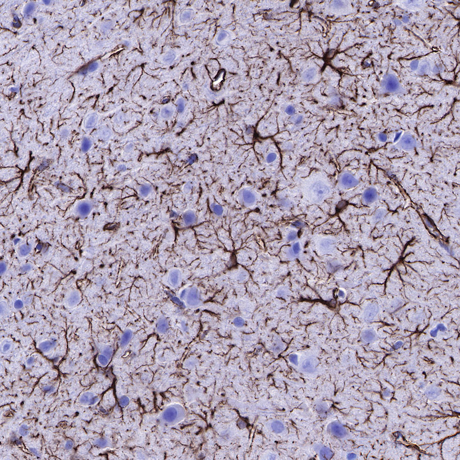
IHC shows positive staining in paraffin-embedded rat cerebral cortex. Anti-GFAP antibody was used at 1/1000 dilution, followed by a HRP Polymer for Mouse & Rabbit IgG (ready to use). Counterstained with hematoxylin. Heat mediated antigen retrieval with Tris/EDTA buffer pH9.0 was performed before commencing with IHC staining protocol.

IHC shows positive staining in paraffin-embedded rat colon. Anti-GFAP antibody was used at 1/1000 dilution, followed by a HRP Polymer for Mouse & Rabbit IgG (ready to use). Counterstained with hematoxylin. Heat mediated antigen retrieval with Tris/EDTA buffer pH9.0 was performed before commencing with IHC staining protocol.
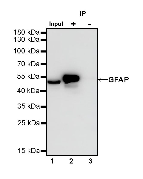
GFAP Rabbit mAb at 1/25 dilution (1 µg) immunoprecipitating GFAP in 0.4 mg rat brain lysate.
Western blot was performed on the immunoprecipitate using GFAP Rabbit mAb at 1/1000 dilution.
Secondary antibody (HRP) for IP was used at 1/400 dilution.
Lane 1: rat brain cell lysate 10 µg (Input)
Lane 2: GFAP Rabbit mAb IP in rat brain lysate
Lane 3: Rabbit monoclonal IgG IP in rat brain lysate
Predicted MW: 50 kDa
Observed MW: 50 kDa
(This blot was developed with high sensitivity substrate)

文献相关图
PFinhibitedRANKL-inducedosteoclastogenesiswithoutcytotoxityinvitro.(A)BMMsweretreatedwith0,2.5,5,and10μMPFinthepresenceof30ng/mL M-CSFand50ng/mLRANKLfor5days.Cellswerefixedwith4%paraformaldehydeandstainedforTRAP.(B)NumberofTRAP-positivecells.(C)AreaofTRAPpositivecells.(D)BMMsweretreatedwith0,2.5,5,and10μMPFinthepresenceof30ng/mLM-CSFfor48,72,and96htomeasurecellviabilitybytheCCK-8 assay.TheresultsshowedthatPFdidnotexhibitcellulartoxicityevenwhentheconcentrationreached40μM.(E)ChemicalstructureofPF,withamolecularformula ofC27H41F2N5Oandamolecularweightof489.64g/mol.Thedataarepresentedasthemean ± SD(*p < 0.05,**p < 0.01,***p < 0.001).
来源于《Open Access Library Journal》
PFimpairedpodosomeactinbeltformationandosteoclastprecursorcellfusion,andinhibitedOCs-mediatedboneresorptionactivityinvitro.(A)BMMswere seeded onto48 well plates with 0,2.5, 5, and 10μM PF in the presence of 30 ng/mL M-CSF and 50 ng/mL RANKL for5 days. Cells were fixed andstained for immunofluorescence.(B)NumberofOCswithF-actinring.(C)AreaofOCswithF-actinring.(D)BMMswereseededontohydroxyapatite-coatedOsteoAssayplates after9-dayculturewithM-CSF(30ng/mL),RANKL(50ng/mL),andwith0,2.5,5,and10μMPF.Representativescanningimagesofboneresorptionpitsareshown. (E)Areaoftheresorptionpits.Thedataarepresentedasthemean ± SD(*p < 0.05,**p < 0.01,***p < 0.001).
来源于《Open Access Library Journal》
PFdepressedtheexpressionofRANKL-inducedosteoclastgenes.BMMsweretreatedwithM-CSF(30ng/mL),RANKL(50ng/mL)inthepresenceof0,2.5,5, and 10 μM PF for 5 days. (A–H) Expression of the osteoclast-specific genes including NFATc1, CTR, CTSK, V-ATPase d2, TRAP and DC-STAMP, and the Notch signalingrelatedgenesincludingNotch2andHes1,wereanalyzedusingquantitativereal-timePCR.RNAexpressionlevelswerenormalizedtotheexpressionof Gapdh.Thedataarepresentedasthemean ± SD(*p < 0.05,**p < 0.01,***p < 0.001).
来源于《Open Access Library Journal》
PFinhibitedosteoclastogenesisbydownregulatingNotch2signalingpathwaysandthephosphorylationofAKT.(A)BMMswereseededat3×105cells/well in6-wellplatesandstimulatedwithM-CSF(30ng/mL)andRANKL(50ng/mL),withorwithoutPF(10μM)for0,1,3and5days.Cellswerelysedandsubjectedto westernblottingusingspecificantibodiesagainstNICD2,Hes1andNFATc1.(B)PFtreatmentsuppressedtheexpressionofNICD2.(C)PFtreatmentsuppressedthe expressionofHes1.(D)PFtreatmentsuppressedtheexpressionofNFATc1.(E)BMMswereseededat5×105cells/wellin6-wellplatesandstimulatedwithRANKL (50 ng/mL), with or without PF (10 μM) for 0, 5, 10, 20, 30, 60s. (F) PF treatment suppressed AKT phosphorylation expression. The data are presented as the mean ± SD(*p < 0.05,**p < 0.01,***p < 0.001).
来源于《Open Access Library Journal》
EffectofPFonosteoblastdifferentiationinvitro.(A,B)MC3T3-E1cellsweretreatedwith0,2.5,5,and10μMPFinthepresenceofascorbicacid(A.A),βglycerophosphate(β-gly)anddexamethasone(DXM)for7,21daysduringosteoblasticdifferentiation.TheALP(Alkalinephosphatase)andAlizarinRedStaining demonstratednosignificantdifferencebetweencontrolandPFtreatedgroups.(C)PFdidnotexhibitcytotoxiceffectonosteoblastprecursorcells.(D)PFdidnot affecttheOBs-relatedgeneexpression.Thedataarepresentedasthemean ± SD(*p < 0.05,**p < 0.01,***p < 0.001).(Forinterpretationofthereferencesto colourinthisfigurelegend,thereaderisreferredtotheWebversionofthisarticle.)
来源于《Open Access Library Journal》
PFsuppressedLPS-inducedcalvarialosteolysisinvivo.(A)Representativeimagesof3Dreconstructionofmicro-CTscanning.(B–D)Bonevolumetotissue volume(BV/TV,%),numberofporosityandthepercentageoftotalporosityofeachsampleweremeasuredandanalyzed.Thedataarepresentedasthemean ± SD (*p < 0.05,**p < 0.01,***p < 0.001).
来源于《Open Access Library Journal》
Histological analysis of the inhibitory effect of PF on LPS-induced bone resorption. (A–C) Representative images of HE and TRAP staining, showing the reducedosteolyticlegionandTRAP-positiveOCsinthePF-treatedgroups.(D–G)RepresentativeimagesofIHCstainingofRANKL,OPG,OCNandTNF-α.(H)TRAPpositiveOCsnumber.(H)QuantitativeanalysisoftheexpressionofRANKL,OPGandRANKL/OPGratio.Thedataarepresentedasthemean ± SD(*p < 0.05, **p < 0.01,***p < 0.001).
来源于《Open Access Library Journal》
温馨提示:本产品仅作科研实验使用
  • 0 购物车
  • 批量
    查询
  • 对比
  • 回顶部