



 专属买赠
专属买赠
 满额赠礼
满额赠礼
| 货号-规格 | 货期 | 价格 | 数量 |
|---|---|---|---|
| abs171703-25uL | 1-2周 | ¥ 600 .00 | |
| abs171703-100uL | 1-2周 | ¥ 2100 .00 | |
| abs171703-1mL | 1-2周 | 询价 |
订购信息:
您可以从我们的授权经销商处购买absin产品或获得技术支持。
实验结果图
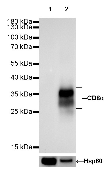
WB result of CD8α Rabbit mAb Primary antibody: CD8α Rabbit mAb at 1/1000 dilution Lane 1: RAW 264.7 whole cell lysate 20 µg Lane 2: mouse thymus lysate 20 µg Negative control: RAW 264.7 whole cell lysate Secondary antibody: #abs20040 at 1/10000 dilution Predicted MW: 29 kDa Observed MW: 28~37 kDa Exposure time: 60s
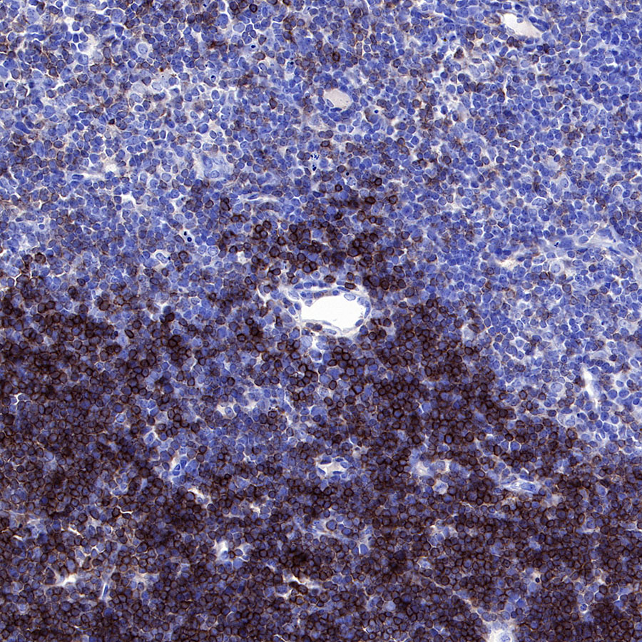
IHC shows positive staining in paraffin-embedded mouse thymus. Anti-CD8α antibody was used at 1/500 dilution, Secondary antibody: #abs20040. Counterstained with hematoxylin. Heat mediated antigen retrieval with Tris/EDTA buffer pH9.0 was performed before commencing with IHC staining protocol.
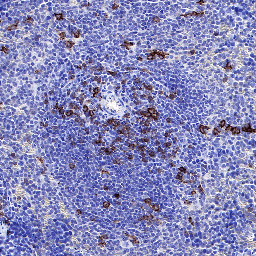
IHC shows positive staining in paraffin-embedded mouse spleen. Anti-CD8α antibody was used at 1/500 dilution, Secondary antibody: #abs20040. Counterstained with hematoxylin. Heat mediated antigen retrieval with Tris/EDTA buffer pH9.0 was performed before commencing with IHC staining protocol.
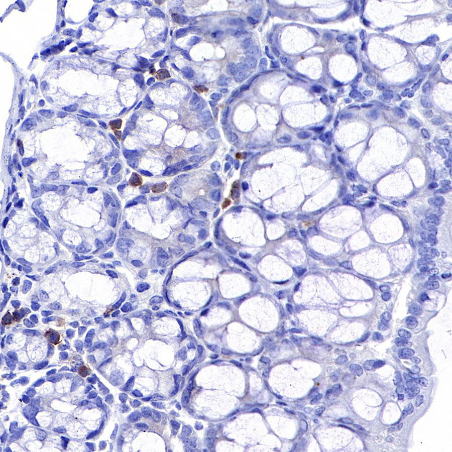
IHC shows positive staining in paraffin-embedded mouse colon. Anti-CD8α antibody was used at 1/500 dilution, Secondary antibody: #abs20040. Counterstained with hematoxylin. Heat mediated antigen retrieval with Tris/EDTA buffer pH9.0 was performed before commencing with IHC staining protocol.
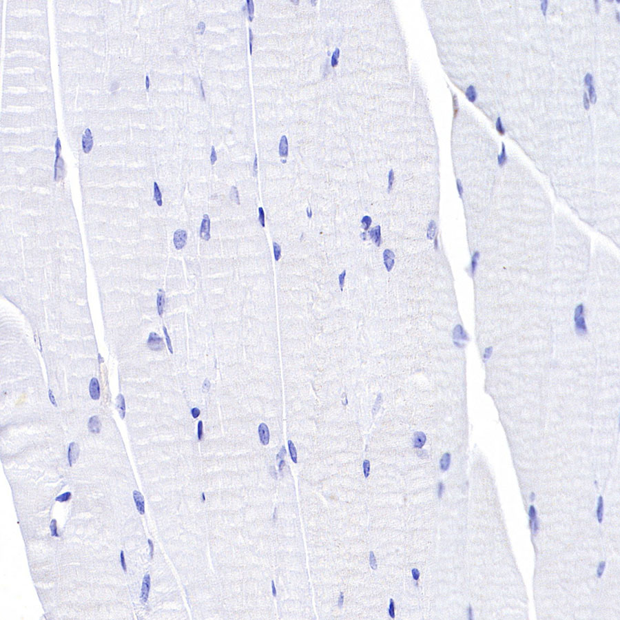
Negative control: IHC shows negative staining in paraffin-embedded mouse cardiac muscle. Anti-CD8α antibody was used at 1/500 dilution, Secondary antibody: #abs20040. Counterstained with hematoxylin. Heat mediated antigen retrieval with Tris/EDTA buffer pH9.0 was performed before commencing with IHC staining protocol.
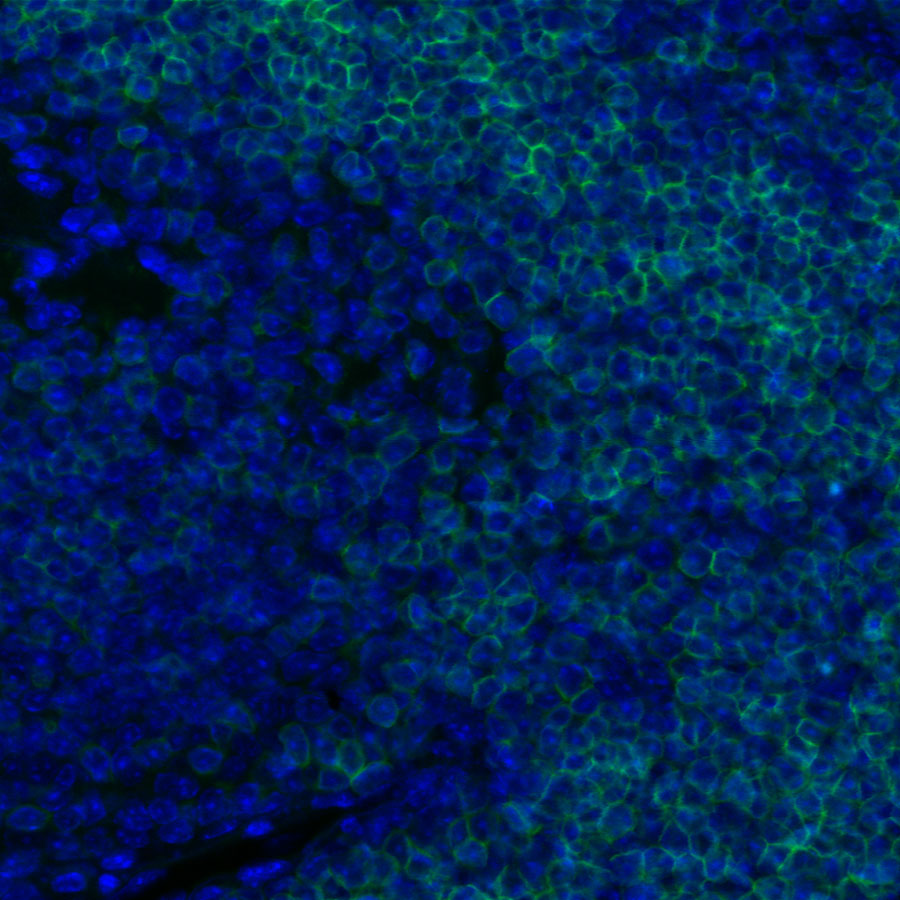
ICC shows positive staining in paraffin-embedded mouse thymus. Anti-CD8α antibody was used at 1/1000 dilution and incubated overnight at 4°C. Secondary antibody: #abs20025 at 1/1000 dilution.Heat mediated antigen retrieval with Tris/EDTA buffer pH9.0 was performed before commencing with ICC staining protocol. Nuclei were counterstained with DAPI.
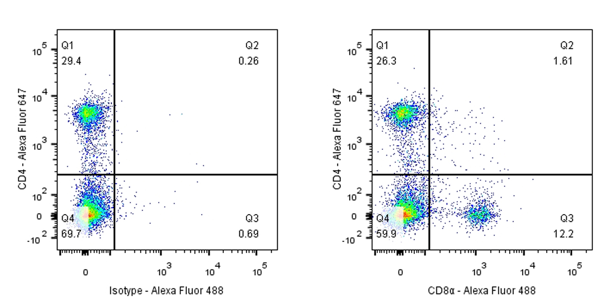
Flow cytometric analysis of mouse primary splenocytes labeling CD8α antibody at 1/50 (1 μg) dilution / (right panel) compared with a Rabbit IgG, Isotype Control / (left panel). Secondary antibody: #abs20025 was used as the secondary antibody. Cells were surface stained with CD4-Alexa Fluor® 647, then stained with rabbit IgG (Left) / CD8α (Right) separately. CD4 and CD8α are mutually exclusive expressed in mouse spleen. Gated on total viable cells.
温馨提示:本产品仅作科研实验使用














 危险品化学品经营许可证(不带存储) 许可证编号:沪(浦)应急管危经许[2022]202585(YS)
  营业执照(三证合一)