



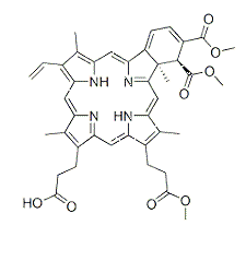
.jpg)
 满额送美团红包
| 货号-规格 | 货期 | 价格 | 数量 |
|---|---|---|---|
| abs817900-5mg | 现货 | ¥ 645 .00 | |
| abs817900-10mg | 2-3天 | ¥ 985 .00 | |
| abs817900-25mg | 2-3天 | ¥ 1995 .00 | |
| abs817900-50mg | 2-3天 | ¥ 2585 .00 |
该货号为 abs47029893 的新货号。
订购信息:
您可以从我们的授权经销商处购买absin产品或获得技术支持。
研究领域
文献相关图

SecretedformofADisintegrinandMetalloproteinase12(ADAM12S)promotestheproliferation,migration,andinvasionofsmallcelllung cancer (SCLC) cells. (A) Green fluorescence and contrast images for H1688 cells stably expressing ZsGreen (control) and ADAM12S/ZsGreen (experiment). Scalebar: 100 μm. (B)Western blotting analysis of cell lysates from H1688 cells stably expressing ZsGreenand ADAM12S/ZsGreen. (C)ADAM12SincreasesthegrowthofH1688cells.TheviabilityofH1688cellsstablyexpressingZsGreenandADAM12S/ZsGreenwasmeasuredby CCK-8 assay. Mean ± SD (n = 4), Student’s t test, *: P < 0.05; **: P < 0.01; ***: P < 0.001. Similar results were obtained from three biological replicates.(D,E)ADAM12SpromotesthemigrationofH1688cells.Woundhealingassay(D)andtranswellmigrationassay(E)wereperformedfor stable H1688 cells. Representative images are provided, and vertical yellow lines indicate the edge of the wound. The migrated distances at eight positions in eachculture well ofthe wound healing assaywere measured. Cells in eight random fields oftranswell chamber membrane were counted. Mean ± SD (n = 8), Student’s t test, ***: P < 0.001. Similar results were obtained from three biological replicates. (F) ADAM12S enhances the invasion of H1688 cells. Matrigel invasion assay was performed for H1688 cells stably expressing ZsGreen and ADAM12S/ZsGreen. Cells in five random fieldsoftranswellchambermembranewerecounted.Mean±SD(n=5),Student’sttest,***:P<0.001.Similarresultswereobtainedfrom three biological replicates.
来源于《J Cell Physiol》

SILACquantitativeproteomicsfortheidentificationofADAM12S-regulatedproteinsinH1688cells.(A)Workflowfortheidentificationof ADAM12S-regulated proteins in H1688 cells. (B) Western blotting analysis of ADAM12S expression in H1688 cells after SILAC labeling. (C) Volcano plot of −Log10 (adjusted P-value) versus Log2 (adjusted H/L ratio) of MS-identified proteins. P values and H/L ratios were calculated by Perseussoftwarefordataobtainedfromthreebiologicalreplicates.Reddots:ADAM12S-regulatedproteins.(D)TenmostenrichedKEGGpathways that ADAM12S-regulated proteins participated in. The categories were obtained from online STRING database analysis.
来源于《J Cell Physiol》

Biochemical validation of the MS-identified ADAM12S-regulated proteins in H1688 cells. (A) Representative Western blotting images of four proteins (ANXA6, CSRP1, HK1, and G6PD) identified by SILAC quantitative proteomics analysis. (B) Quantification of Western blotting imagesforANXA6,CSRP1,HK1,andG6PDfromthreebiologicalreplicates.Mean±SD(n=3),Student’sttest,*:P<0.05;**:P<0.01;***:P< 0.001.
来源于《J Cell Physiol》

Proteomics and biochemical analyses reveal that ADAM12S upregulates HK1 in SCLC cells. (A) HK1 tryptic peptides identified by the quantitativeproteomicsanalysis.The aminoacidsequence,chargestate(z),MH+, meanH/Lratio,andMaxQuantscore areshown inthetable.(B) Representative tandem MS spectrum of an HK1 tryptic peptide. The peptide sequence, MH+, m/ z, charge state (z), and Δmass are depicted in the annotatedspectrogram.(C)MSspectrumofthecorrespondingHK1trypticpeptidefrompanelB.Thepeptidesequence,MH+,chargestate(z),m/z, Δmass,andH/Lratioaredepicted.m/zoftheisotopepeaksfromthelightandheavyamino-acid-codedsamplesarelabeled.(D)qPCRtomeasurethe relative mRNA levels of ADAM12S after siRNA knockdown. GAPDH was used for normalization. Mean ± SD (n = 4), Student’s t test, *: P < 0.05. Similarresultswereobtainedfromthreebiologicalreplicates.(E)ADAM12knockdownreducestheHK1proteinlevelinH446cells.H446cellswere transfected with two siADAM12s, and the resulting cell lysates were immunoblotted. (F) Quantification of HK1 from three biological replicates of Western blotting analyses in panel E. Mean ± SD (n = 3), Student’s t test, *: P < 0.05; **: P < 0.01.
来源于《J Cell Physiol》

ADAM12SpromotestheproliferationandmetastasisofSCLCcellsviaHK1.(A)WesternblottinganalysistoverifythesiHK1knockdown efficiency in H1688 cells. Stable H1688 cell lines were transfected with control and HK1 siRNA, and the resulting cell lysates were used for immunoblotting analysis. (B) HK1 knockdown partially inhibits the ADAM12S-mediated increase in the proliferation of H1688 cells. CCK-8 assay wasused tomeasure the cell proliferation. Mean±SD (n = 4), Student’s ttest (at 96 h),***: P <0.001; ns: not significant. (C,D) HK1 knockdown completely abolishes the effect of ADAM12S on the migration (C) and invasion (D) of H1688 cells. Cells in eight random fields of the transwell chamberwerecounted. Mean±SD (n=8), Student’s ttest,**:P<0.01;***:P< 0.001;ns: notsignificant. Similar results wereobtainedfromtwo biological replicates. (E) Western blotting analysis to verify the shHK1 knockdown efficiency of H1688 cells. H1688 cells stably expressing ZsGreen andADAM12S/ZsGreenwereinfectedwithshCtrlorshHK1lentiviralparticlesandselectedbypuromycinfor2weeks.Thecelllysatesweresubjected toimmunoblottinganalysis.(F)HK1knockdowncompletelyeliminatestheeffectofADAM12SonthecolonyformationofH1688cells.Mean±SD (n = 4), Student’s t test, ***: P < 0.001; ns: not significant. Similar results were obtained from two biological replicates.
来源于《J Cell Physiol》

HK1 knockdown attenuates the ADAM12S-mediated elevation of glucose consumption and lactate production. (A) qPCR measurementoftherelativemRNAlevelsofglycolysis-relatedgenesin H1688 cells stably expressing control and ADAM12S. GAPDH was usedfornormalization.Mean±SD(n=4),Student’sttest,*:P<0.05; **:P<0.01;***:P<0.001;ns:notsignificant.(B,C)Relativeglucose consumption (B) and lactate production (C) in H1688 cells with ADAM12S expression and/or HK1 knockdown. Mean ± SD (n ≥ 3), Student’s t test, *: P < 0.05; **: P < 0.01; ns: not significant. Similar results were obtained from three biological replicates.
来源于《J Cell Physiol》

Proposed model for the regulation of metabolic pathways by ADAM12S in SCLC cells. ADAM12S upregulates HK1 and enhances HK1-dependent aerobic glycolysis, thereby promoting the proliferation, migration, and invasion of SCLC cells.
来源于《J Cell Physiol》
温馨提示:本产品仅作科研实验使用














 危险品化学品经营许可证(不带存储) 许可证编号:沪(浦)应急管危经许[2022]202585(YS)
  营业执照(三证合一)