



SUZ12 抗体;ChET 9 protein antibody; CHET9 antibody; Chromatin precipitated E2F target 9 protein antibody; JJAZ1 antibody; Joined to JAZF1 protein antibody; KIAA0160 antibody; Polycomb protein SUZ12 antibody; Suppressor of zeste 12 homolog antibody; Suppressor of zeste 12 protein homolog antibody; SUZ12 antibody; SUZ12 polycomb repressive complex 2 subunit antibody; SUZ12_HUMAN antibody
| 货号-规格 | 货期 | 价格 | 数量 |
|---|---|---|---|
| abs155784-50ug | 1-2周 | ¥ 1500 .00 | |
| abs155784-100ug | 1-2周 | ¥ 2300 .00 |
订购信息:
您可以从我们的授权经销商处购买absin产品或获得技术支持。若要查看您所在地区的经销商,请从以下的下拉列表中选择。
研究领域
实验结果图
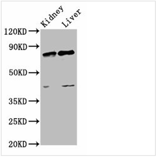
Western Blot
Positive WB detected in: Rat kidney tissue, Rat liver tissue
All lanes: SUZ12 antibody at 3.4μg/ml
Secondary
Goat polyclonal to rabbit IgG at 1/50000 dilution
Predicted band size: 84 kDa
Observed band size: 84 kDa
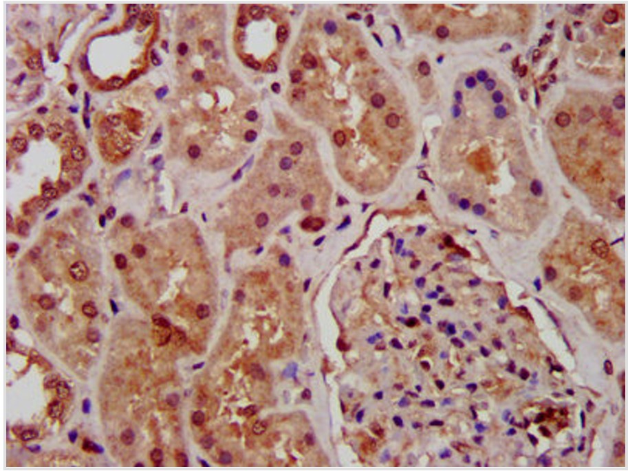
IHC image of abs155784 diluted at 1:1000 and staining in paraffin-embedded human kidney tissue performed on a Leica BondTM system. After dewaxing and hydration, antigen retrieval was mediated by high pressure in a citrate buffer (pH 6.0). Section was blocked with 10% normal goat serum 30min at RT. Then primary antibody (1% BSA) was incubated at 4°C overnight. The primary is detected by a biotinylated secondary antibody and visualized using an HRP conjugated SP system.
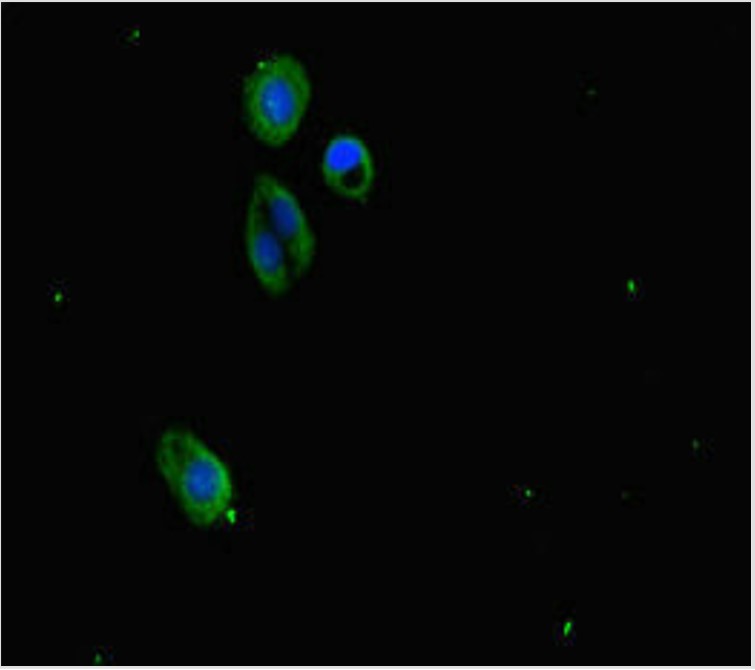
Immunofluorescence staining of HepG2 cells with abs155784 at 1:333, counter-stained with DAPI. The cells were fixed in 4% formaldehyde, permeabilized using 0.2% Triton X-100 and blocked in 10% normal Goat Serum. The cells were then incubated with the antibody overnight at 4°C. The secondary antibody was Alexa Fluor 488-congugated AffiniPure Goat Anti-Rabbit IgG(H+L).
温馨提示:本产品仅作科研实验使用


 危险品化学品经营许可证(不带存储) 许可证编号:沪(浦)应急管危经许[2022]202585(YS)
  营业执照(三证合一)