



 专属买赠
专属买赠
 满额赠礼
满额赠礼

| 货号-规格 | 货期 | 价格 | 数量 |
|---|---|---|---|
| abs955-50T | 现货 | ¥ 2625 .00 |
订购信息:
您可以从我们的授权经销商处购买absin产品或获得技术支持。
实验结果图
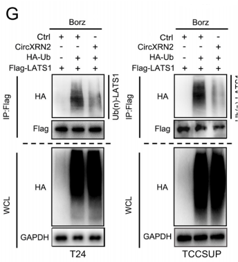
检测不同细胞中LATS1蛋白质的泛素化情况。如图所示,在硼替佐米和N-乙基马来酰亚胺预处理8小时(防止蛋白降解,防止去泛素化)。通过免疫沉淀实验检测对照组和circXRN2过表达组中泛素化的LATS1蛋白水平,结果表明circXRN2过表达细胞中的LATS1蛋白较少被泛素化。Xie B, Lin J, Chen X, Zhou X, Zhang Y, Fan M, Xiang J, He N, Hu Z, Wang F. CircXRN2 suppresses tumor progression driven by histone lactylation through activating the Hippo pathway in human bladder cancer. Mol Cancer. 2023 Sep 8;22(1):151. doi: 10.1186/s12943-023-01856-1. PMID: 37684641; PMCID: PMC10486081.

利用IP-MS检测与LATS1相互作用的蛋白质,其中SPOP是一种E3连接酶。Xie B, Lin J, Chen X, Zhou X, Zhang Y, Fan M, Xiang J, He N, Hu Z, Wang F. CircXRN2 suppresses tumor progression driven by histone lactylation through activating the Hippo pathway in human bladder cancer. Mol Cancer. 2023 Sep 8;22(1):151. doi: 10.1186/s12943-023-01856-1. PMID: 37684641; PMCID: PMC10486081.

免疫共沉淀结果显示,野生型LATS1可以与SPOP结合,但SBC1突变体LATS1与SPOP的相互作用几乎完全消失(n)。敲低circXRN2促进了相互作用(o)。Xie B, Lin J, Chen X, Zhou X, Zhang Y, Fan M, Xiang J, He N, Hu Z, Wang F. CircXRN2 suppresses tumor progression driven by histone lactylation through activating the Hippo pathway in human bladder cancer. Mol Cancer. 2023 Sep 8;22(1):151. doi: 10.1186/s12943-023-01856-1. PMID: 37684641; PMCID: PMC10486081.
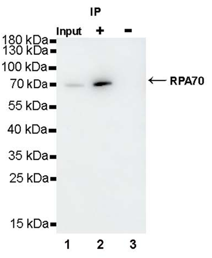
Lane 1: HeLa whole cell lysate 10 μg (lnput)
Lane 2: RPA70 Recombinant Rabbit mAb IP in HeLa whole cell lysate
Lane 3: Rabbit monoclonal lgG IP in HeLa whole cell lysate
文献相关图
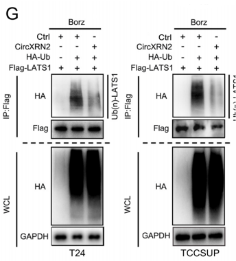
检测不同细胞中LATS1蛋白质的泛素化情况。如图所示,在硼替佐米和N-乙基马来酰亚胺预处理8小时(防止蛋白降解,防止去泛素化)。通过免疫沉淀实验检测对照组和circXRN2过表达组中泛素化的LATS1蛋白水平,结果表明circXRN2过表达细胞中的LATS1蛋白较少被泛素化。
来源于《Molecular Cancer》 DOI:10.1186/s12943-023-01856-1
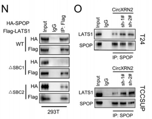
免疫共沉淀结果显示,野生型LATS1可以与SPOP结合,但SBC1突变体LATS1与SPOP的相互作用几乎完全消失(n)。敲低circXRN2促进了相互作用(o)。
来源于《Molecular Cancer》 DOI:10.1186/s12943-023-01856-1
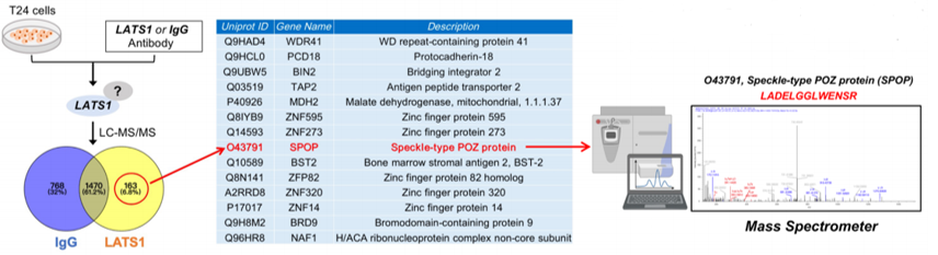
利用IP-MS检测与LATS1相互作用的蛋白质,其中SPOP是一种E3连接酶。
来源于《Molecular Cancer》 DOI:10.1186/s12943-023-01856-1
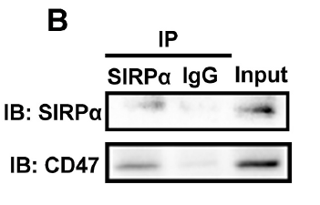
来源于《Bioact Mater》 DOI:10.1016/j.bioactmat.2025.01.012
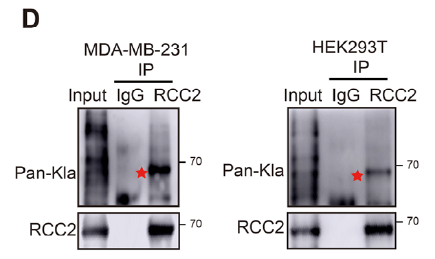
Detection of endogenous lactylation of RCC2 protein inMDAMB-231 and HEK293T cells
来源于《Adv Sci (Weinh)》 DOI:10.1002/advs.202415530
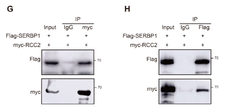
G,H) CoIP and WB analysis of RCC2 and SERBP1
来源于《Adv Sci (Weinh)》 DOI:10.1002/advs.202415530
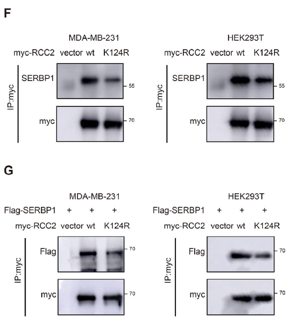
F,G) Endogenous and exogenous IP and WB analysis of the interaction intensity between RCC2 and SERBP1 when overexpressed with wt-RCC2 or mut-RCC2.
来源于《Adv Sci (Weinh)》 DOI:10.1002/advs.202415530
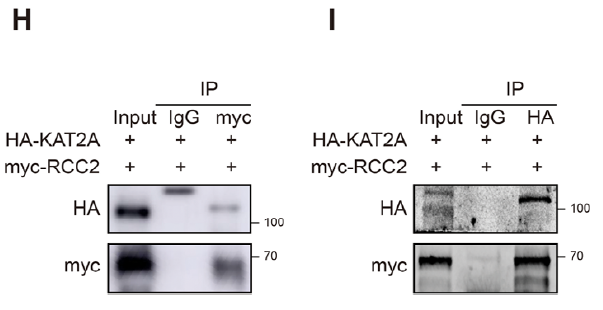
H,I) CoIP and WB analysis of RCC2 and KAT2A.
来源于《Adv Sci (Weinh)》 DOI:10.1002/advs.202415530
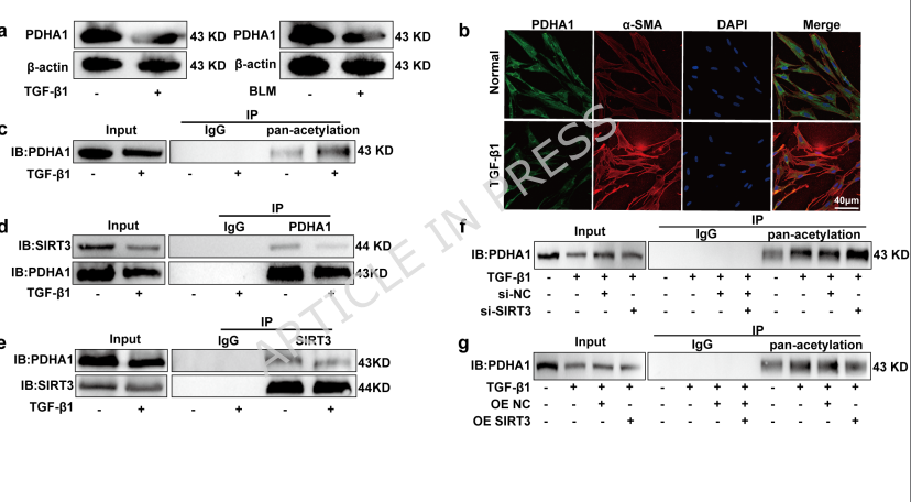
(C) Immunoblot analysis of PDHA1 acetylation in TGF-β1-stimulated fibroblasts following immunoprecipitation. (D) Co-IP assay using an anti-PDHA1 antibody for immunoprecipitation in MRC-5 cells with or without TGF-β1 stimulation, followed by immunoblotting with anti-SIRT3 and anti-PDHA1 antibodies. The “Input” panels show the expression levels of SIRT3 and PDHA1 in whole cell lysates, demonstrating SIRT3 downregulation. (E) Reverse Co-IP assay using an anti-SIRT3 antibody for immunoprecipitation, followed by immunoblotting with anti-PDHA1 and anti-SIRT3 antibodies. Input panels confirm SIRT3 downregulation. (F) MRC-5 cells were transfected with control or SIRT3-targeting siRNA. PDHA1 acetylation was assessed by immunoprecipitation with a pan-acetylation antibody followed by immunoblotting with an anti-PDHA1 antibody. (G) MRC-5 cells were transfected with a vector control (OE NC) or a SIRT3-overexpressing (OE SIRT3) plasmid. PDHA1 acetylation was assessed as in (C).
来源于《Cellular and Molecular Life Sciences》 DOI:10.1007/s00018-026-06166-5
温馨提示:本产品仅作科研实验使用















 危险品化学品经营许可证(不带存储) 许可证编号:沪(浦)应急管危经许[2022]202585(YS)
  营业执照(三证合一)