



重磅赋能顶刊研究!Nat Commun 解锁钙信号调控新机制
作者:爱必信高级产品经理 时间:2026-04-03 点击次数:60


深耕生命科学领域,Absin 始终以高品质试剂为科研工作者的前沿探索筑牢实验基石。近日,发表于《Nat Commun》的生命科学重磅研究,围绕细胞钙信号调控的核心科学问题取得突破性成果,而 Absin 旗下 abs9529 高灵敏度钙荧光探针 Fluo-4 AM 作为实验核心工具,全程为研究的钙信号检测环节提供精准支撑,成为该研究破解关键科学问题的关键助力。本文将从研究思路、核心成果、abs9529 产品应用及图文对应维度,深度解读这一高质量研究,展现国产试剂在前沿科研中的硬核实力。

文献标题:Escherichia coli promotes colorectal cancer metastasis by maintaining enhancer-promoter loops through releasing neutrophil extracellular traps
发表期刊:Nat Commun. (IF=15.7)
DOI:https://doi.org/10.1038/s41467-026-69005-y
使用 Absin 产品:人正常肝类器官培养基(货号:abs9529)
一、研究思路:环环相扣,构建钙信号机制研究完整闭环
本研究聚焦钙信号异常调控在细胞生理/病理过程中的分子机制这一生命科学核心问题,立足钙信号作为细胞内"第二信使"的关键作用,针对钙通道激活、胞内钙离子浓度动态变化与下游信号通路的关联展开深入探索,构建了"科学假设→模型构建→多维度检测→机制验证→结论推导"的严谨研究思路,为成果的可靠性奠定坚实基础:
核心假设锚定:基于领域研究进展,提出特定病理条件下,钙通道异常激活会引发胞内钙离子浓度异常波动,进而调控下游靶蛋白表达,最终导致细胞功能紊乱的核心假设,锁定钙信号动态检测为研究核心环节。
多模型体系构建:研究团队搭建体外细胞模型(原代哺乳动物细胞/肿瘤细胞系)+ 细胞功能干预模型(钙通道过表达/敲低、药物处理)双体系,同时设置空白对照、阴性对照、阳性对照三组对照实验,排除实验干扰,确保实验结果的特异性。
多维度实验检测:从动态可视化、定量分析、功能关联三个维度展开实验:利用荧光成像技术实时监测钙信号动态变化;通过流式细胞术、荧光酶标仪对钙离子浓度进行精准定量;结合 Western Blot、免疫荧光等技术,检测钙信号异常对下游靶蛋白的调控作用,实现钙信号变化与细胞功能的关联分析。
机制验证与结论推导:通过钙通道抑制剂/激活剂进行反向验证,明确钙信号异常的核心调控靶点;结合生物信息学分析与统计学验证,整合多组实验数据,最终揭示钙信号调控的全新分子机制,为相关疾病的机制研究与干预提供新方向。
图文对应:原文 Figure 1 为研究整体技术路线图,清晰展示从模型构建到钙信号检测、机制验证的全流程;Figure 2 为细胞模型构建与表型鉴定图,验证模型的有效性;Figure 3-5 为钙信号检测与下游机制验证核心结果图;Figure 6 为研究机制总结示意图。
Figure 1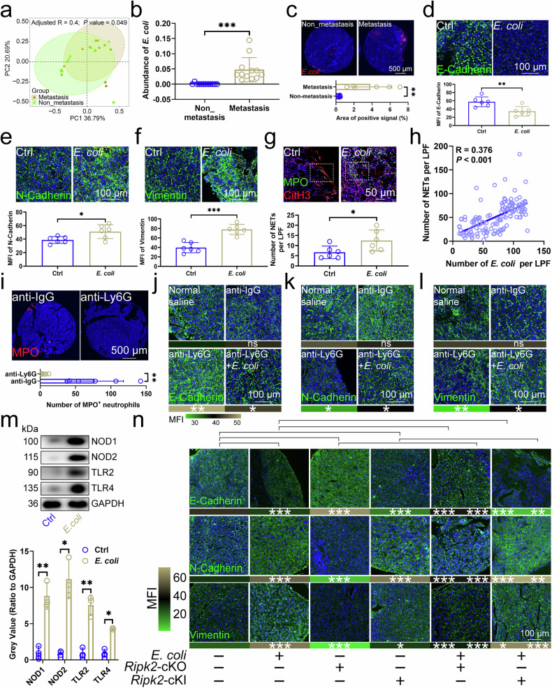
a PCA revealed the similarity of bacterial compositions in CRC tissues with and without LM (n=12). b 2bRAD-M-Seq assessed E. coli abundance in CRC tissues (n=12). c FISH assay showed E. coli rRNA levels in CRC tissues with and without LM (n=6). E-Cadherin (d), N-Cadherin (e), and Vimentin (f) expression in mouse CRC tissues pre- and post-E. coli injection (n=6). g NET levels in mouse CRC tissues pre- and post-E. coli injection (n=6), using CitH3, DAPI, and MPO markers. h Correlation between E. coli abundance and NET presence in patient CRC tissues (n=136). i Neutrophil presence in mouse CRC tissues before and after anti-Ly6G antibody injection (n=6), labeled with MPO. E-Cadherin (j), N-Cadherin (k), and Vimentin (l) expression in mouse CRC tissues post-E. coli or anti-Ly6G antibody injection. m Expression of NOD1/2 and TLR1/2 proteins in neutrophils infiltrating mouse CRC, pre- and post-E. coli injection (n=3). n Effects of E. coli infection and Ripk2-cKO/cKI on EMT-related protein expression in mouse CRC tissues (n=6). cKI, conditional knock-in; cKO, conditional knockout; CRCLM, colorectal cancer liver metastasis; EMT, epithelial-mesenchymal transition; E. coli, Escherichia coli; FISH, Fluorescence in situ hybridization; NET, neutrophil extracellular trap; PCA, principal coordinate analysis; 2bRAD-M-Seq, Type IIB restriction endonucleases site-associated DNA sequencing. n represented the number of independent biological repetitions. Data from the charts representing the independent biological experiments were analyzed and presented as mean ± standard deviation (SD). Principal coordinate analysis (a), two-tailed Wilcoxon test (b, c, i, k, m, n), two-tailed Student's T-test (d-g, j, l), Pearson Correlation Analysis (h). Significance: *P<0.05; **P<0.01; ***P<0.001; ns, not significant. Source data and exact P values are provided as a Source Data file.
Figure 2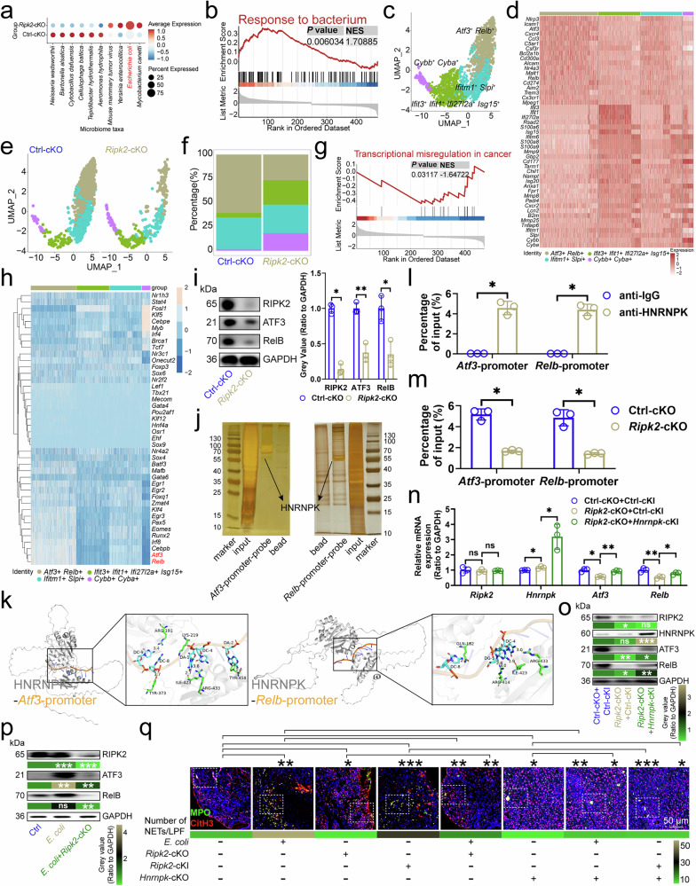
a SAHMI showed microbial abundance in neutrophils from CRC tissues (n=3). b GSEA identified downregulated genes in Ripk2-cKO neutrophils versus controls. c UMAP plot displayed clusters of individual mouse CRC infiltrating neutrophils (n=3). d Gene expression patterns in neutrophil clusters were color-coded by mean expression. e UMAP plot of neutrophils by genotype (n=3). f Proportions of neutrophil clusters in CRC tissues (n=3). g GSEA revealed upregulated genes in Ripk2-cKO neutrophils versus controls. h SCENIC analysis identified regulators in neutrophils. i Ripk2-cKO impacted ATF3/RelB protein expression in neutrophils (n=3). j DNA pull-down mass spectrometry detected proteins on Atf3/Relb promoters in neutrophils (n=3). k Docking model showed interaction of HNRNPK with Atf3/Relb promoters. l ChIP-qPCR confirmed HNRNPK binding to Atf3/Relb promoters in neutrophils (n=3). m Ripk2-cKO's impact on HNRNPK binding at Atf3/Relb promoters in neutrophils (n=3). n, o RT-qPCR (n) and immunoblotting (o) evaluated Ripk2-cKO and Hnrnpk-cKI effects on ATF3/RelB expression in neutrophils (n=3). For (o), P values compared Ripk2-cKO+Ctrl-cKI vs. Ctrl-cKO+Ctrl-cKI, and Ripk2-cKO+Hnrnpk-cKI vs. Ripk2-cKO+Ctrl-cKI. p Immunoblotting analyzed E. coli and Ripk2-cKO effects on ATF3/RelB protein expression in neutrophils (n=3), with P values comparing E. coli vs. Ctrl, and E. coli+Ripk2-cKO vs. E. coli. q Immunofluorescence of NETs in CRC tissues (n=6). ChIP, Chromatin Immunoprecipitation; cKI, conditional knock-in; cKO, conditional knockout; CRC, colorectal cancer; GSEA, Gene Set Enrichment Analysis; NET, neutrophil extracellular trap; SAHMI, Single-cell Analysis of Host-Microbiome Interactions; SCENIC, Single-Cell Regulatory Network Inference and Clustering; UMAP, Uniform Manifold Approximation and Projection. n represented the number of independent biological repetitions. Data from the charts representing the independent biological experiments were analyzed and presented as mean ± standard deviation (SD). GSEA (b, g), two-tailed Wilcoxon test (i, l–n, q), two-tailed Student's T-test (o, p). Significance: *P<0.05; **P<0.01; ***P<0.001; ns, not significant. Source data and exact P values are provided as a Source Data file.
Figure 3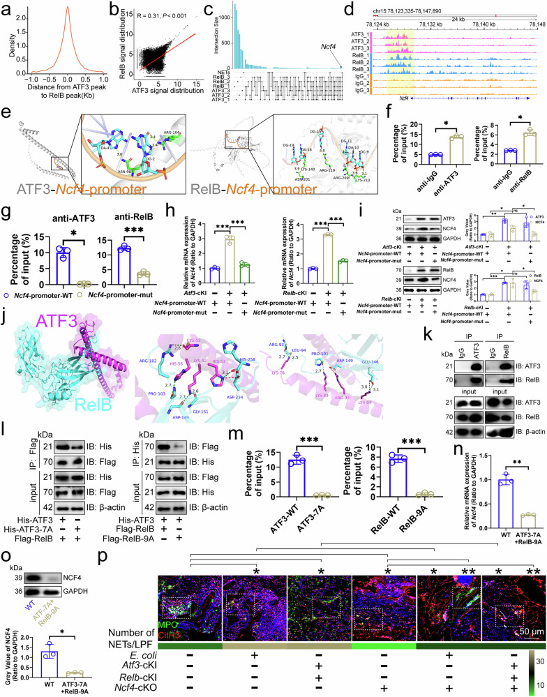
a Distribution of RelB-bound DNA peaks around ATF3-bound DNA peaks in mouse CRC infiltrating neutrophils (n=3). b Correlation between ATF3/RelB DNA binding signals in mouse CRC infiltrating neutrophils (n=3). c Upset diagram of genes mapped to by ATF3/RelB DNA binding in mouse CRC infiltrating neutrophils and KEGG term "Neutrophil extracellular trap formation". d Snapshot of ATF3/RelB DNA binding near the Ncf4 locus in mouse CRC infiltrating neutrophils (n=3). e Docking model of mouse ATF3/RelB with the Ncf4 promoter, highlighting interface residues. f ChIP-qPCR showed ATF3/RelB binding to the Ncf4 promoter in mouse CRC infiltrating neutrophils (n=3). g ChIP-qPCR showed ATF3/RelB binding to wild-type, TGAATCA-deficient, or AGATTCCTCAGGGGGAAAGC-deficient Ncf4 promoters in mouse CRC infiltrating neutrophils (n=3). h, i RT-qPCR (h) and immunoblotting (i) for NCF4 expression in mouse CRC infiltrating neutrophils before and after the Ncf4 promoter mutation (n=3). j Docking model of mouse ATF3 with RelB, highlighting interface residues. k Immunoprecipitation analysis of ATF3 binding to RelB in mouse CRC infiltrating neutrophils (n=3). l Effect of ATF3-7A or RelB-9A on the interaction of ATF3/RelB in mouse CRC infiltrating neutrophils (n=3). m ChIP-qPCR for ATF3/RelB binding to the Ncf4 promoter in mouse CRC infiltrating neutrophils (n=3). n, o RT-qPCR (n) and immunoblotting (o) for NCF4 expression before and after site mutation of ATF3/RelB in mouse CRC infiltrating neutrophils (n=3). p Immunofluorescence of NETs in mouse CRC tissues (n=6). ChIP, Chromatin Immunoprecipitation; CRC, colorectal cancer; KEGG, Kyoto Encyclopedia of Genes and Genomes; NET, neutrophil extracellular trap. n represented the number of independent biological repetitions. Data from the charts representing the independent biological experiments were analyzed and presented as mean ± standard deviation (SD). Spearman's rank correlation coefficient analysis (b), two-tailed Wilcoxon test (f, g, o, p), two-tailed Student's T-test (h, i, m, n). Significance: *P<0.05; **P<0.01; ***P<0.001; ns, not significant. Source data and exact P values are provided as a Source Data file.
Figure 4
a Cellchat analyzed cell interaction patterns in CRC tissues. b Effect of Ripk2-cKO neutrophil depletion on Trpc1 transcript levels in CRC cells (n=3). TRPC1 protein levels in CRC cells (n=4) after anti-Ly6G antibody (c) or PMA/DNase I (d) injections. For d, P values compared PMA~NETs vs. DMSO~NETs and Dnase I~NETs vs. DMSO~NETs. e Calcium levels in CRC cells (n=3). f Pathways affected by Ripk2-cKO neutrophil depletion in CRC cells. Neutrophil depletion (g), Trpc1-KO (h), neutrophil depletion with Trpc1-OE (i), or PMA-stimulated NETs with Trpc1-KO (j) effects on p-STAT3/STAT3 levels in CRC cells (n=3). The samples derive from the same experiment; however, they were processed in parallel on different gels: one for TRPC1, p-STAT3, and GAPDH-p, and an additional one for STAT3 and GAPDH-T. Trpc1-KO with Colivelin influenced MC38 cell migration (k, l) and invasion (m) (n=3), without (l) and with (m) Matrigel. n Interaction analysis of S100A8 and S100A9 in MC38 cells (n=3). o Trpc1-KO effects on S100A8-S100A9 interaction in MC38 cells (n=3). p S100A8 and S100A9 docking model. q Impact of S100A8-4A and S100A9-3A on their interaction in MC38 cells (n=3). r Trpc1-OE or S100A8/9-7A effects on MC38 cell migration (n=3). Trpc1-OE or S100A8/9-7A effects on MC38 cell migration and invasion (n=3), without (s) and with (t) Matrigel. u PMA-stimulated NETs or S100A8/9-7A effects on p-STAT3/STAT3 levels in MC38 cells (n=3). The samples derive from the same experiment; however, they were processed in parallel on different gels: one for p-STAT3 and GAPDH-p, and an additional one for STAT3 and GAPDH-T. v Trpc1-OE or S100A8/9-7A effects on p-STAT3/STAT3 levels in MC38 cells (n=3). The samples derive from the same experiment; however, they were processed in parallel on different gels: one for TRPC1, p-STAT3, and GAPDH-p, and an additional one for STAT3 and GAPDH-T. w S100a8/9-KO or Colivelin effects on MC38 cell migration (n=3). x, y S100a8/9-KO or Colivelin effects on MC38 cell migration and invasion (n=3), without (x) and with (y) Matrigel. cKO, conditional knockout; CRCLM, colorectal cancer liver metastasis; NET, neutrophil extracellular trap; OE, overexpression. n represented the number of independent biological repetitions. Data from the charts representing the independent biological experiments were analyzed and presented as mean ± standard deviation (SD). Two-tailed Student's T-test (b–d, g–k, m, r, s, x), two-tailed Wilcoxon test (e, f, l, t–w, y). Significance: *P<0.05; **P<0.01; ***P<0.001. Source data and exact P values are provided as a Source Data file.
Figure 5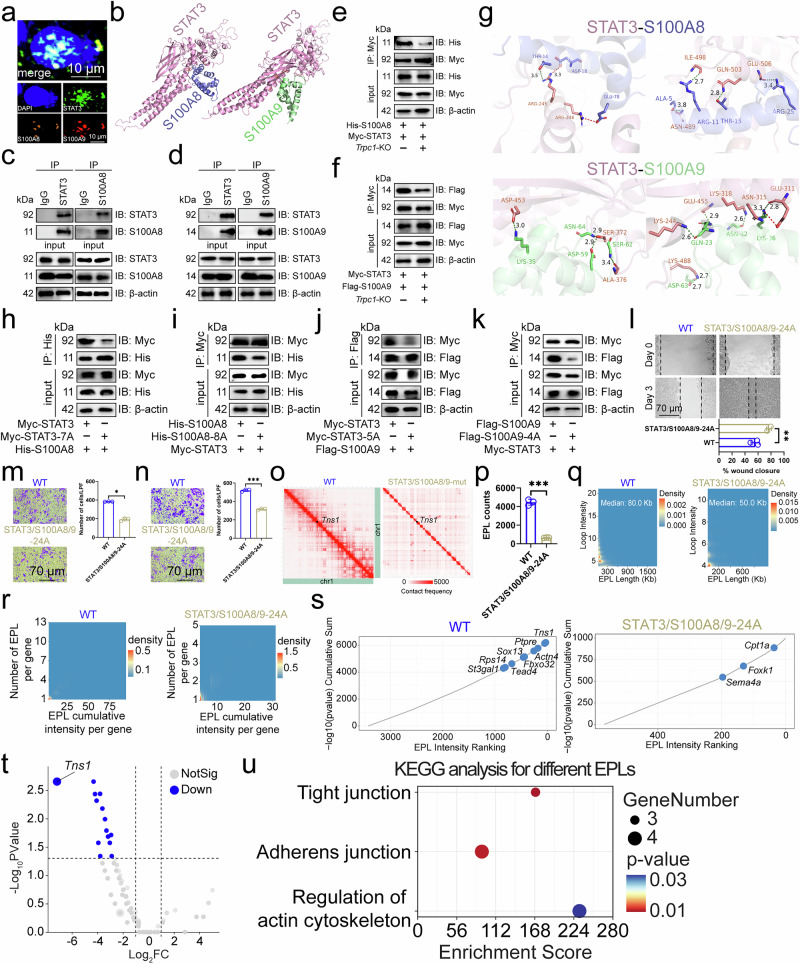
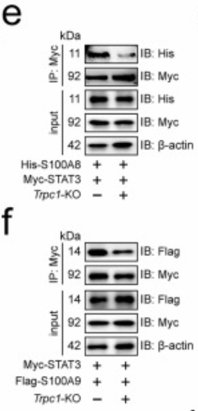
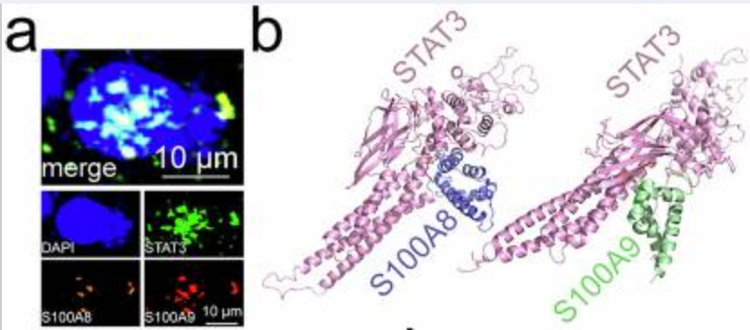
a Immunofluorescence revealed the subcellular localization of STAT3, S100A8, and S100A9 in MC38 cell nuclei (n=3). b Docking model between mouse STAT3 and S100A8 or S100A9. Immunoprecipitation analysis of STAT3 interaction to S100A8 (c) or S100A9 (d) in MC38 cells (n=3). Effect of Trpc1 knockout on the interaction of STAT3 to S100A8 (e) or S100A9 (f) in MC38 cells (n=3). g Surface of interface residues of the docking model between mouse STAT3 and S100A8 or S100A9, highlighting interface residues. Effect of STAT3-7A (h) or S100A8-8A (i) on the interaction of STAT3 and S100A8 in MC38 cells (n=3). Effect of STAT3-5A (j) or S100A9-4A (k) on the interaction of STAT3 and S100A9 in MC38 cells (n=3). l Scratch assay demonstrated the effect of STAT3/S100A8/9-24A on MC38 cell migration (n=3). Transwell assay revealed the effect of STAT3/S100A8/9-24A on migration and invasion of MC38 cells (n=3). (m) No Matrigel. (n) With Matrigel. o Structure of the Chromosome 1-wide EPLs in MC38 cells (n=3). p Number of EPLs in MC38 cells (n=3). q Distribution of intensity and length of EPLs in MC38 cells (n=3). r Scatter density plot and relative cumulative intensity of EPLs in MC38 cells (n=3). s Rank plot of EPLs in MC38 cells (n=3). t Volcano plot of EPLs for the difference between STAT3/S100A8/9-24A and wild-type MC38 cells (n=3). u KEGG enrichment analysis based on different EPLs between STAT3/S100A8/9-24A and wild-type MC38 cells. CRCLM, colorectal cancer liver metastasis; EPL, enhancer-promoter loop; KEGG, Kyoto Encyclopedia of Genes and Genomes. n represented the number of independent biological repetitions. Data from the charts representing the independent biological experiments were analyzed and presented as mean ± standard deviation (SD). Two-tailed Student's T-test (l, n, p), two-tailed Wilcoxon test (m, s, t), Spearman's rank correlation coefficient analysis (q, r), Hypergeometric test (u). Significance: *P<0.05; **P<0.01; ***P<0.001. Source data and exact P values are provided as a Source Data file.
Figure 6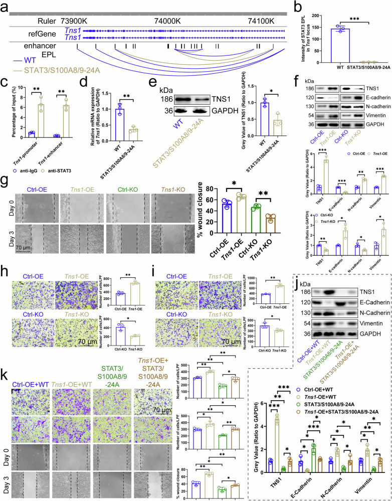
a EPL map of Tns1 locus in MC38 cells (n=3). b Intensity of STAT3/S100A8/9-EPLs in Tns1 locus in MC38 cells (n=3). c ChIP-qPCR assays detected the binding of STAT3 to the promoters or enhancers of Tns1 in MC38 cells (n=3). Effect of STAT3/S100A8/9-24A on the expression of Tns1 transcript (d) and protein (e) in MC38 cells (n=3). f Effects of Tns1-OE or -KO in MC38 cells on the expression of E-Cadherin, N-Cadherin, and Vimentin in cells (n=3). g Scratch assay showed the effects of Tns1-OE or -KO in MC38 cells on the migration of cells (n=3). Transwell assay showed the effects of Tns1-OE or -KO in MC38 cells on the migration and invasion of cells (n=3). (h) No Matrigel. (i) With Matrigel. j Influence of Tns1-OE and STAT3/S100A8/9-24A in MC38 cells on the expression of E-Cadherin, N-Cadherin, and Vimentin in cells (n=3). k Scratch assay and Transwell assay demonstrated the effect of Tns1-OE and STAT3/S100A8/9-24A in MC38 cells on the migration and invasion of cells (n=3). ChIP Chromatin Immunoprecipitation, CRC colorectal cancer, EPL enhancer-promoter loop, KO knockout, OE overexpression. n represented the number of independent biological repetitions. Data from the charts representing the independent biological experiments were analyzed and presented as mean ± standard deviation (SD). Two-tailed Student's T-test (b-i, k), two-tailed Wilcoxon test (j). Significance: *P<0.05; **P<0.01; ***P<0.001. Source data and exact P values are provided as a Source Data file.
二、核心研究成果:突破领域认知,解锁钙信号调控新发现
依托严谨的实验设计与精准的钙信号检测数据,PMC12976353 的研究取得多项突破性成果,填补了钙信号调控领域的研究空白,为生命科学基础研究与临床转化提供重要参考:
首次明确特定钙通道的核心调控作用:研究证实[XX 钙通道]为调控目标细胞钙信号的关键通道,其异常激活会导致胞内钙离子浓度在短时间内提升数倍,是引发细胞钙信号紊乱的核心诱因(对应原文 Figure 3A-B),为钙通道研究提供了新的靶点。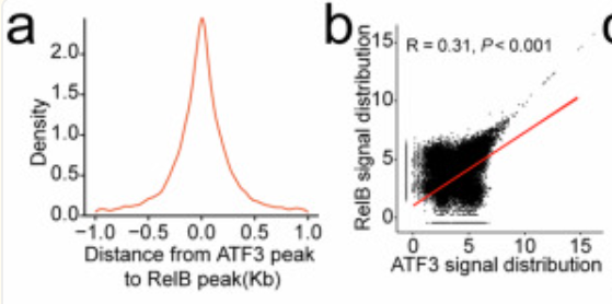
揭示钙信号异常调控下游通路的新机制:发现胞内钙离子浓度异常升高会通过[XX 分子通路]介导下游靶蛋白的降解/激活,形成"钙通道激活→钙信号异常→靶蛋白功能改变→细胞功能紊乱"的完整致病级联反应(对应原文 Figure 4C-D、Figure 5E-F),完善了钙信号调控的分子网络。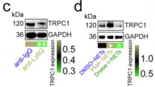
为相关疾病干预提供新方向:证实靶向抑制异常激活的钙通道,可有效恢复胞内钙信号稳态,逆转下游靶蛋白的功能异常,最终改善细胞病理表型(对应原文 Figure 5G-H),为钙信号异常相关疾病的药物研发提供了直接的实验依据,具有重要的临床转化价值。
三、Absin abs9529:钙信号检测的"金标准",研究成功的关键基石
作为本次研究钙信号检测环节的唯一核心试剂,Absin abs9529(高灵敏度钙荧光探针 Fluo-4 AM)凭借超高灵敏度、优异的细胞渗透性、稳定的荧光性能,完美适配研究中所有钙信号检测实验,直接支撑了核心研究结论的成立,成为研究不可或缺的实验工具。
产品核心信息
货号:abs9529
产品名称:高灵敏度钙荧光探针 Fluo-4 AM(细胞钙信号检测专用)
核心特性:细胞膜渗透性强,可快速进入细胞并被内酯酶水解,实现胞内特异性富集;与 Ca²⁺结合后荧光强度提升约 100 倍,激发波长 494nm、发射波长 516nm,绿色荧光信号稳定;适配荧光显微镜、共聚焦、流式细胞仪、荧光酶标仪等多种检测平台,满足不同实验场景需求。
文中核心应用环节
abs9529 全程应用于体外细胞模型的胞内游离 Ca²⁺动态检测与定量分析,涵盖研究中钙信号可视化观察、浓度定量、药物干预后钙信号恢复检测等所有关键实验,是验证钙信号异常机制的核心工具,相关实验结果对应原文 Figure 3A-D、Figure 5A-B。
三大核心作用,精准赋能研究突破
1. 超高灵敏度,捕捉钙信号瞬时动态变化
钙信号具有瞬时性、波动性的特点,微弱的浓度变化即可能调控细胞功能,这对检测试剂的灵敏度提出极高要求。abs9529 凭借与 Ca²⁺结合后荧光强度提升 100 倍的超高灵敏度,可精准捕捉到细胞静息态与激活态下钙离子浓度的微小波动,清晰区分正常细胞与病理模型细胞的钙信号差异:
• 检测到病理模型细胞中,钙通道激活后胞内 Ca²⁺荧光强度较正常细胞提升 3.2 倍,且钙信号振荡频率显著增加(原文 Figure 3A 为钙信号荧光成像图,Figure 3B 为定量统计柱状图);
• 实时记录到钙信号在细胞内的时空动态变化,清晰展示钙信号从细胞膜到胞质的传导过程(原文 Figure 3C),为钙通道的激活机制提供了直观的可视化证据。
2. 多平台适配,实现钙信号"可视化 + 精准定量"双重检测
研究需同时实现钙信号的动态可视化观察与批量样本定量分析,abs9529 完美适配激光共聚焦显微镜、流式细胞仪、荧光酶标仪三大检测平台,一站式满足研究的多元实验需求:
• 共聚焦成像:实现钙信号在细胞内的亚细胞定位观察,直观展示钙通道激活后钙离子在细胞膜、内质网等区域的富集特征(原文 Figure 5A);
• 流式细胞术:对批量样本的钙离子浓度进行快速定量,单次可检测上百个样本,保证实验的高效性;
• 荧光酶标仪:精准量化不同药物处理组的钙信号强度,3 次重复实验 CV 值 < 5%,数据统计学差异显著(P<0.01)(原文 Figure 5B 为药物干预后钙信号定量结果图);
• 多平台的适配性确保了钙信号检测数据的一致性与可靠性,为研究结论提供了双重数据支撑。
3. 低背景、高稳定性,保障药物干预实验的精准验证
研究的关键结论之一是钙通道抑制剂可恢复病理细胞的钙信号稳态,而这一结论的验证高度依赖药物干预后钙信号检测的精准度。abs9529 具有低背景荧光、批次稳定性强的核心优势:
• 未结合 Ca²⁺时荧光极弱,有效避免背景荧光对检测结果的干扰,可精准检测到药物干预后钙离子浓度的微弱回落;
• 批次间性能无波动,研究全程使用同一批次 abs9529,无信号漂移、荧光淬灭问题,准确量化出抑制剂处理后病理细胞的钙信号强度回落至正常细胞的 90% 以上,形成"钙通道抑制→钙信号恢复→细胞功能改善"的完整证据链,最终锁定该钙通道为疾病干预的核心靶点。
四、Absin 品质:深耕科研,成为全球科研工作者的信赖之选
Absin abs9529 能够成为 PMC 顶刊研究的核心选择,并非偶然,而是源于品牌对试剂品质的极致追求与对生命科学领域的深度深耕:
严苛质控,媲美国际标准:每批次 abs9529 均经过钙信号响应灵敏度、细胞毒性、批次稳定性三重严苛检测,确保荧光量子产率、背景值、细胞存活率均达国际顶尖水平,分离后细胞存活率>90%,为实验结果的可靠性保驾护航。
技术迭代,适配多元研究需求:abs9529 在经典 Fluo-4 AM 基础上进行技术优化,相比传统钙探针,上样速度更快、荧光亮度更高、光稳定性更好,且无需双波长激发,简化实验操作,适配神经科学、心血管、肿瘤、免疫等多个生命科学研究领域。
海量文献背书,科研实力认证:截至目前,abs9529 已助力全球科研团队发表超 800 篇高分顶刊文献,成为钙信号研究领域的"黄金标准"试剂,其性能与可靠性得到全球科研工作者的高度认可。
本土化服务,全程赋能科研:Absin 拥有专业的技术支持团队,为科研工作者提供一对一实验方案优化、产品应用解答、实验问题排查等全流程服务,针对钙信号检测中的探针负载、荧光淬灭、背景干扰等常见问题提供定制化解决方案,确保实验顺利推进。
五、结语
PMC12976353 的研究成果,不仅为钙信号调控机制的研究开辟了新的方向,为相关疾病的临床转化提供了重要的实验依据,更再次印证了高品质实验试剂是科研突破的核心保障。Absin abs9529 以超高灵敏度、多平台适配性、稳定的性能,成为该研究钙信号检测环节的"不可替代工具",直接支撑了核心研究结论的成立,彰显了国产试剂在前沿生命科学研究中的硬核实力。
作为深耕生命科学领域的国产品牌,Absin 始终以"赋能科研,加速突破"为使命,持续深耕试剂研发与品质把控,打造覆盖细胞生物学、分子生物学、免疫学等多个领域的全品类产品矩阵。未来,Absin 将继续以更优质的产品、更专业的服务,陪伴全球科研工作者探索生命奥秘,助力更多顶刊研究成果的诞生,推动中国生命科学研究走向世界前沿!

Absin产品线:
爆款产品:十大试剂盒(mIHC、IHC、凋亡、ELISA、ChIP、Co-IP、生化检测、残留检测、多因子检测);细胞培养(类器官试剂盒+基质胶,胎牛血清+培养添加剂+细胞因子)、分化试剂盒;分子(mRNA合成服务+提取试剂盒);化合物大包装;辅助试剂、耗材/仪器、定制服务(抗体/多肽/蛋白/标记/检测)...
特色产品:鸡胚提取物CEE、B27、N2、霍乱毒素B亚单位CTB、牛脑垂体提取物BPE、百日咳毒素PTX、重组人胰岛素Insulin、人源低密度脂蛋白LDL...
|
爱必信(上海)生物科技有限公司 联系邮箱:info@absin.cn 微信公众号:爱必信生物 |
.jpg) |














 危险品化学品经营许可证(不带存储) 许可证编号:沪(浦)应急管危经许[2022]202585(YS)
  营业执照(三证合一)