一大波抗体来袭——经组化验证,病理研究适用
时间:2022-10-24 点击次数:187
分享:


|
|
|
Absin 抗体正在火热促销中,限时7折! 活动截止至2022年12月31日 |

|
货号 |
名称 |
反应种属 |
应用 |
|
Rabbit anti-c-Met mAb (009-7) |
Human |
WB;IHC-P |
|
|
Rabbit anti-CLDN1 mAb (025-43) |
Human,Mouse |
IHC-P;ICC |
|
|
Rabbit anti-GCDFP-15 mAb(010-579) |
Human |
WB;IHC-P |
|
|
Rabbit anti-Keratin 14 mAb (023-12) |
Human,Mouse |
WB;IHC-P;ICC |
|
|
Rabbit anti-Keratin 8 mAb (016-46) |
Human |
WB;IHC-P;ICC |
|
|
Rabbit-CD31 mAb (008-17) |
Human |
WB;IHC-P |

WB result of c-Met Rabbit mAb at 1/5000 dilution Lane 1 : Hela whole cell lysate 20 µg Lane 2 : HT-29 whole cell lysate 20 µg Lane 3 : SK-OV-3 whole cell lysate 20 µg Secondary antibody: #abs20040 at 1/10000 dilution Predicted MW: 155 kDa Observed MW: 155 kDa Exposure time: 9 seconds.
IHC shows positive staining in paraffin-embedded human colon. Anti-c-Met antibody was used at 1/250 dilution, Secondary antibody: #abs20040 Counterstained with hematoxylin. Heat mediated antigen retrieval with Tris/EDTA buffer pH9.0 was performed before commencing with IHC staining protocol.
IHC shows positive staining in paraffin-embedded human cervix cancer. Anti-c-Met antibody was used at 1/250 dilution, Secondary antibody: #abs20040 Counterstained with hematoxylin. Heat mediated antigen retrieval with Tris/EDTA buffer pH9.0 was performed before commencing with IHC staining protocol.
Rabbit anti-CLDN1 mAb (025-43)
IHC shows membrane staining in paraffin-embedded human tonsil squamous epithelium. Anti-CLDN1 antibody was used at 1/250 dilution, Secondary antibody: #abs20040 Counterstained with hematoxylin. Heat mediated antigen retrieval with Tris/EDTA buffer pH9.0 was performed before commencing with IHC staining protocol.
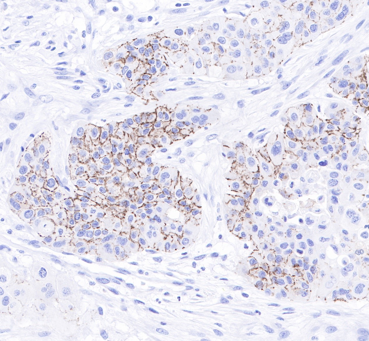
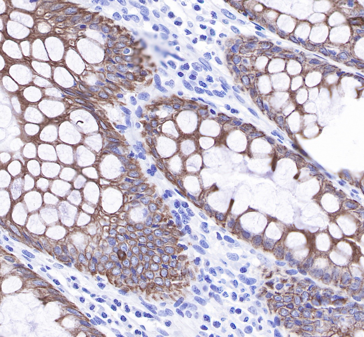
IHC shows membrane staining in paraffin-embedded human pancreatic cancer. Anti-CLDN1 antibody was used at 1/250 dilution, Secondary antibody: #abs20040. Counterstained with hematoxylin. Heat mediated antigen retrieval with Tris/EDTA buffer pH9.0 was performed before commencing with IHC staining protocol.
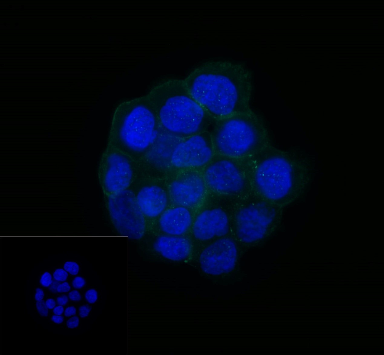
ICC shows membrane staining in A431 cells. Anti-CLDN1 antibody was used at 1/250 dilution and incubated overnight at 4°C. Secondary antibody: #abs20025 The cells were fixed with 100% methanol and permeabilized with 0.1% PBS-Triton X-100. Nuclei were countersained with DAPI.
IHC shows membrane staining in paraffin-embedded mosue colon. Anti-CLDN1 antibody was used at 1/250 dilution, Secondary antibody: #abs20040. Counterstained with hematoxylin. Heat mediated antigen retrieval with Tris/EDTA buffer pH9.0 was performed before commencing with IHC staining protocol.
Rabbit anti-GCDFP-15 mAb(010-579)
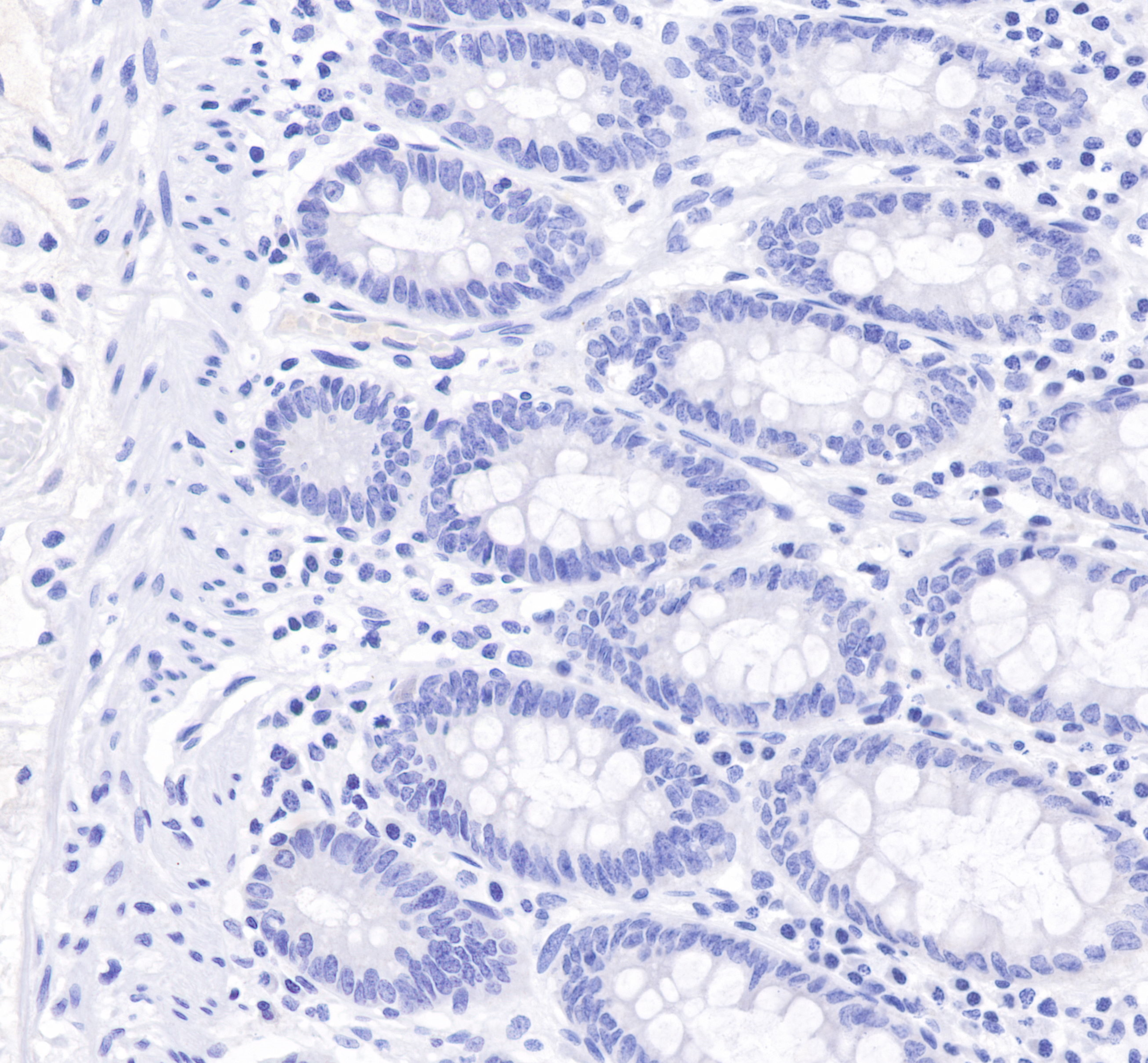
IHC shows negative staining in paraffin-embedded human colon (negative tissue). Anti-GCDFP-15 antibody was used at 1/250 dilution, Secondary antibody: #abs20040. Counterstained with hematoxylin.Heat mediated antigen retrieval with Tris/EDTA buffer pH9.0 was performed before commencing with IHC staining protocol.
Rabbit anti-Keratin 14 mAb (023-12)
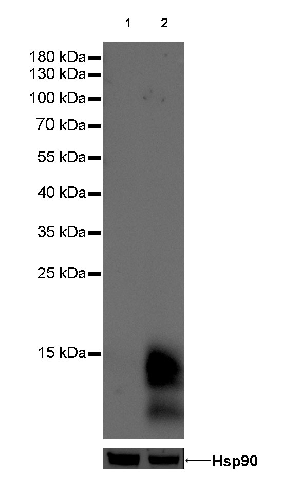
 WB result of Keratin 14 Rabbit mAb at 1/10000 dilution Lane 1 : A431 whole cell lysate 10 µg Secondary antibody:#abs20040 at 1/10000 dilution Predicted MW: 51 kDa Observed MW: 51 kDa Exposure time: 2 seconds.
 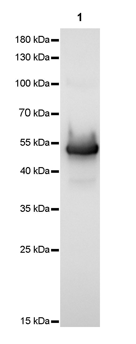 WB result of Keratin 14 Rabbit mAb at 1/5000 dilution Lane 1 : mouse skin lysate 10 µg Secondary antibody: #abs20040 at 1/10000 dilution Predicted MW: 51 kDa Observed MW: 51 kDa Exposure time: 2 seconds.
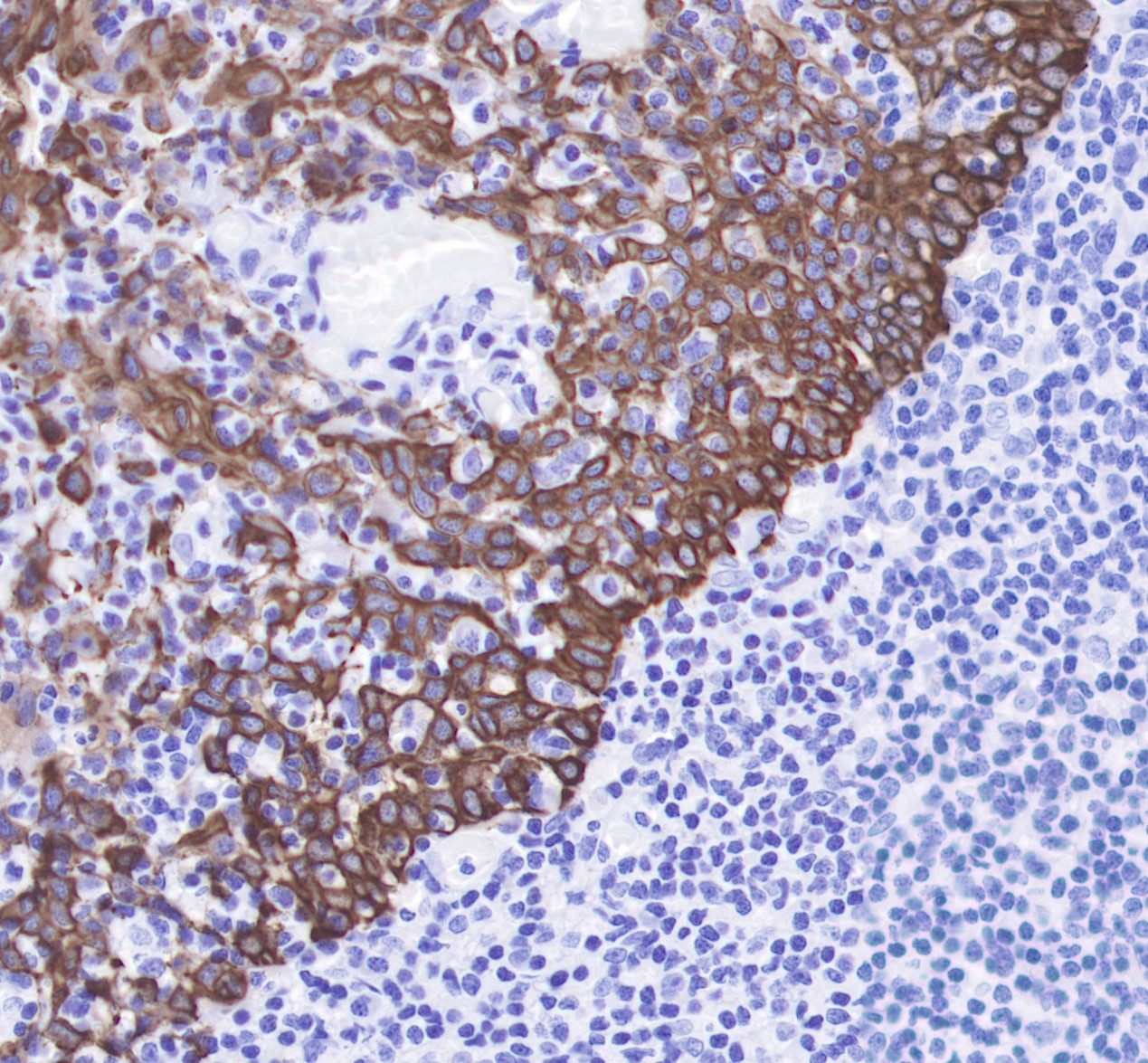
IHC shows membrane staining in paraffin-embedded human tonsil squamous epithelium. Anti-Keratin 14 antibody was used at 1/4000 dilution, Secondary antibody: #abs20040 Counterstained with hematoxylin. Heat mediated antigen retrieval with Tris/EDTA buffer pH9.0 was performed before commencing with IHC staining protocol.
ICC shows membrane staining in A431 cells. Anti-Keratin 14 antibody was used at 1/500 dilution and incubated overnight at 4°C. Secondary antibody:#abs20025 The cells were fixed with 100% methanol and permeabilized with 0.1% PBS-Triton X-100. Nuclei were countersained with DAPI.
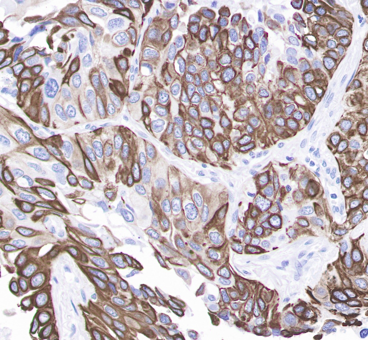
IHC shows membrane staining in paraffin-embedded human lung squamous cell cancer. Anti-Keratin 14 antibody was used at 1/4000 dilution, Secondary antibody: #abs20040 Counterstained with hematoxylin. Heat mediated antigen retrieval with Tris/EDTA buffer pH9.0 was performed before commencing with IHC staining protocol.
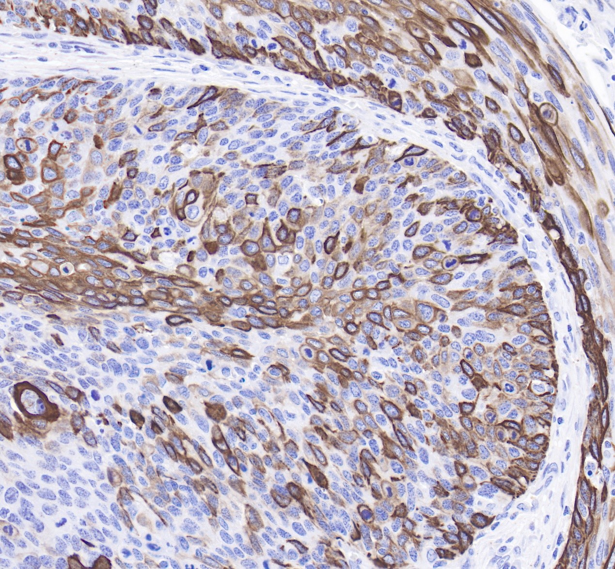
IHC shows membrane staining in paraffin-embedded human cervix cancer. Anti-Keratin 14 antibody was used at 1/4000 dilution, Secondary antibody: #abs20040 Counterstained with hematoxylin. Heat mediated antigen retrieval with Tris/EDTA buffer pH9.0 was performed before commencing with IHC staining protocol.
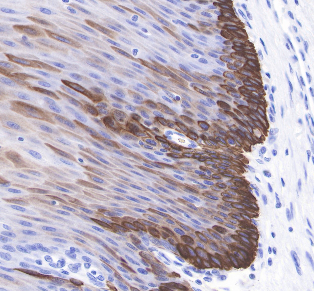
IHC shows membrane staining in paraffin-embedded human esophagus. Anti-Keratin 14 antibody was used at 1/4000 dilution, Secondary antibody: #abs20040 Counterstained with hematoxylin. Heat mediated antigen retrieval with Tris/EDTA buffer pH9.0 was performed before commencing with IHC staining protocol.
Rabbit anti-Keratin 8 mAb (016-46)
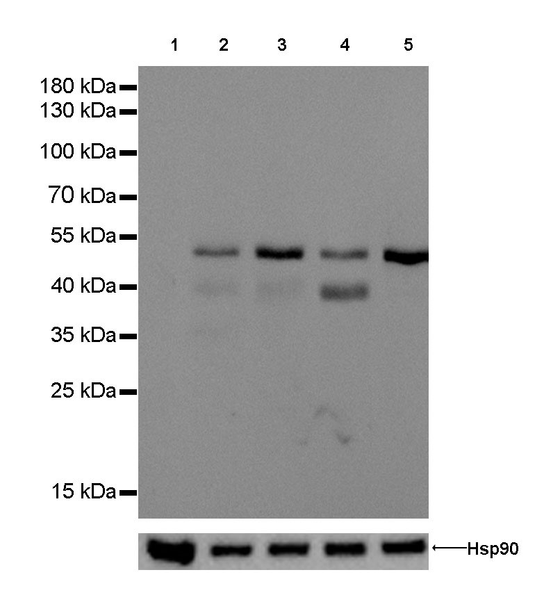
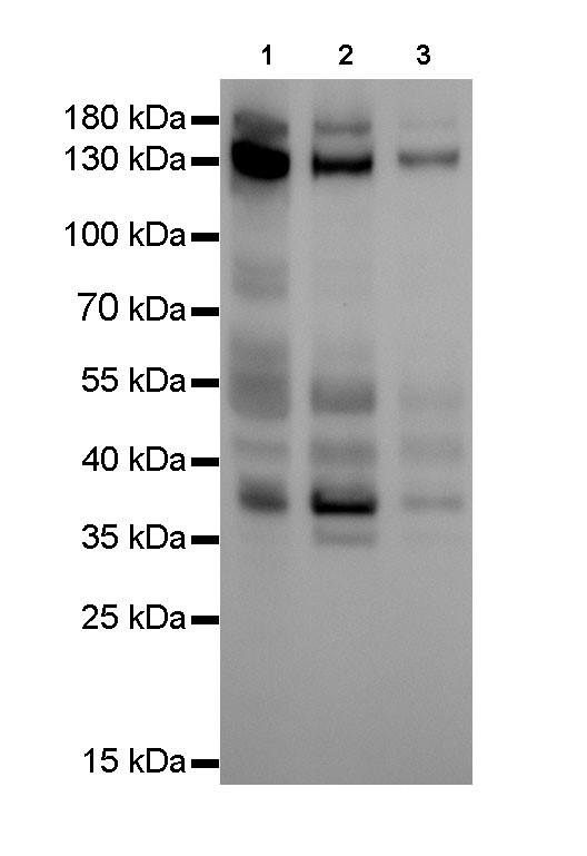
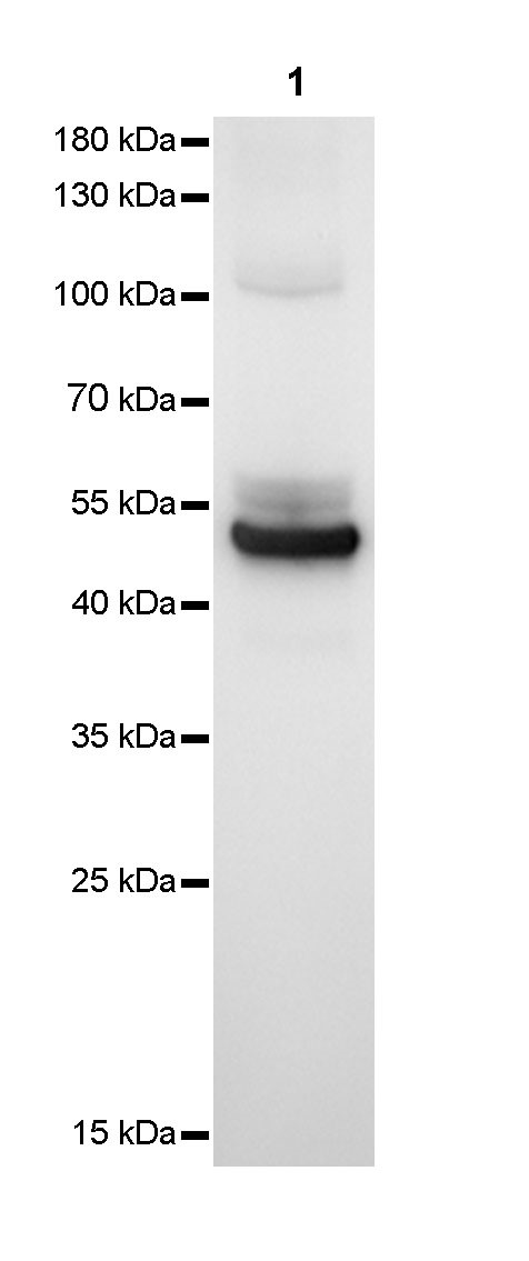
WB result of anti-Keratin 8 antibody at 1/1000 dilution Lane 1 : Jurkat whole cell lysate 20 µg Lane 2 : A549 whole cell lysate 20 µg Lane 3 : SK-BR-3 whole cell lysate 20 µg Lane 4 : MCF-7 whole cell lysate 20 µg Lane 5 : Hela whole cell lysate 20 µg Negative control: Jurkat whole cell lysate Secondary antibody: #abs20040 at 1/10000 dilution Predicted MW: 55 kDa Observed MW: 53 kDa Exposure time: 120 seconds.
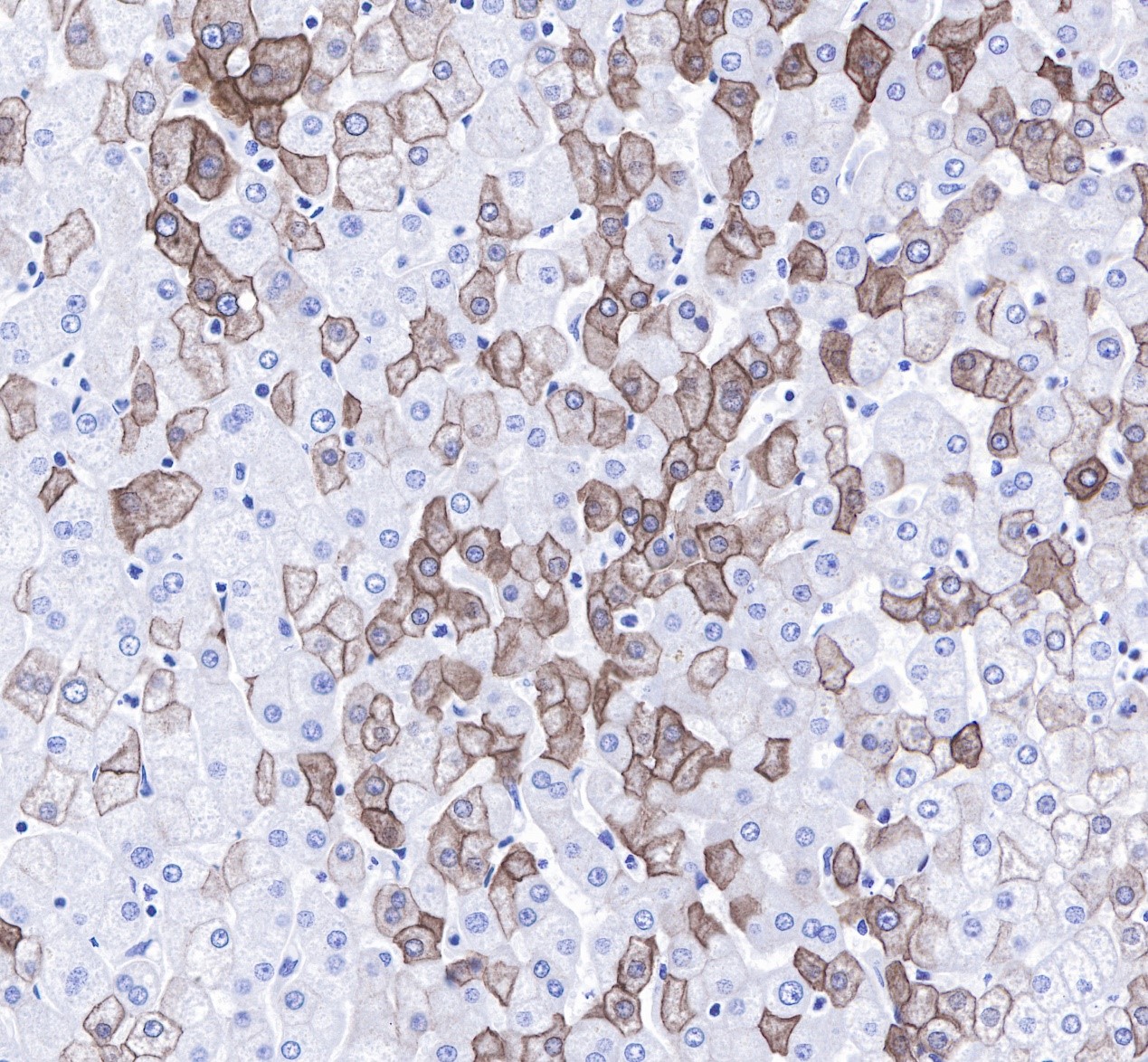
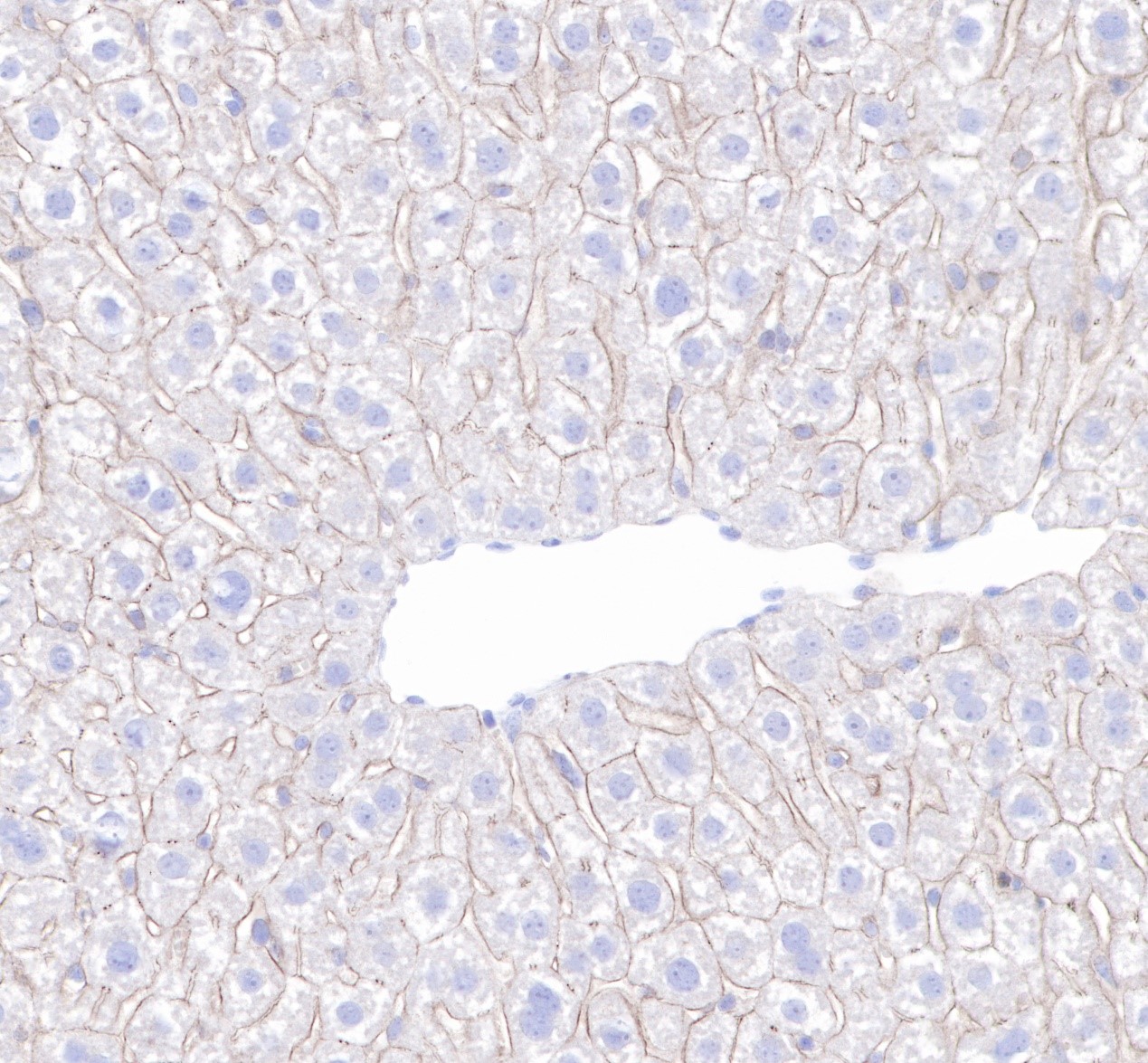
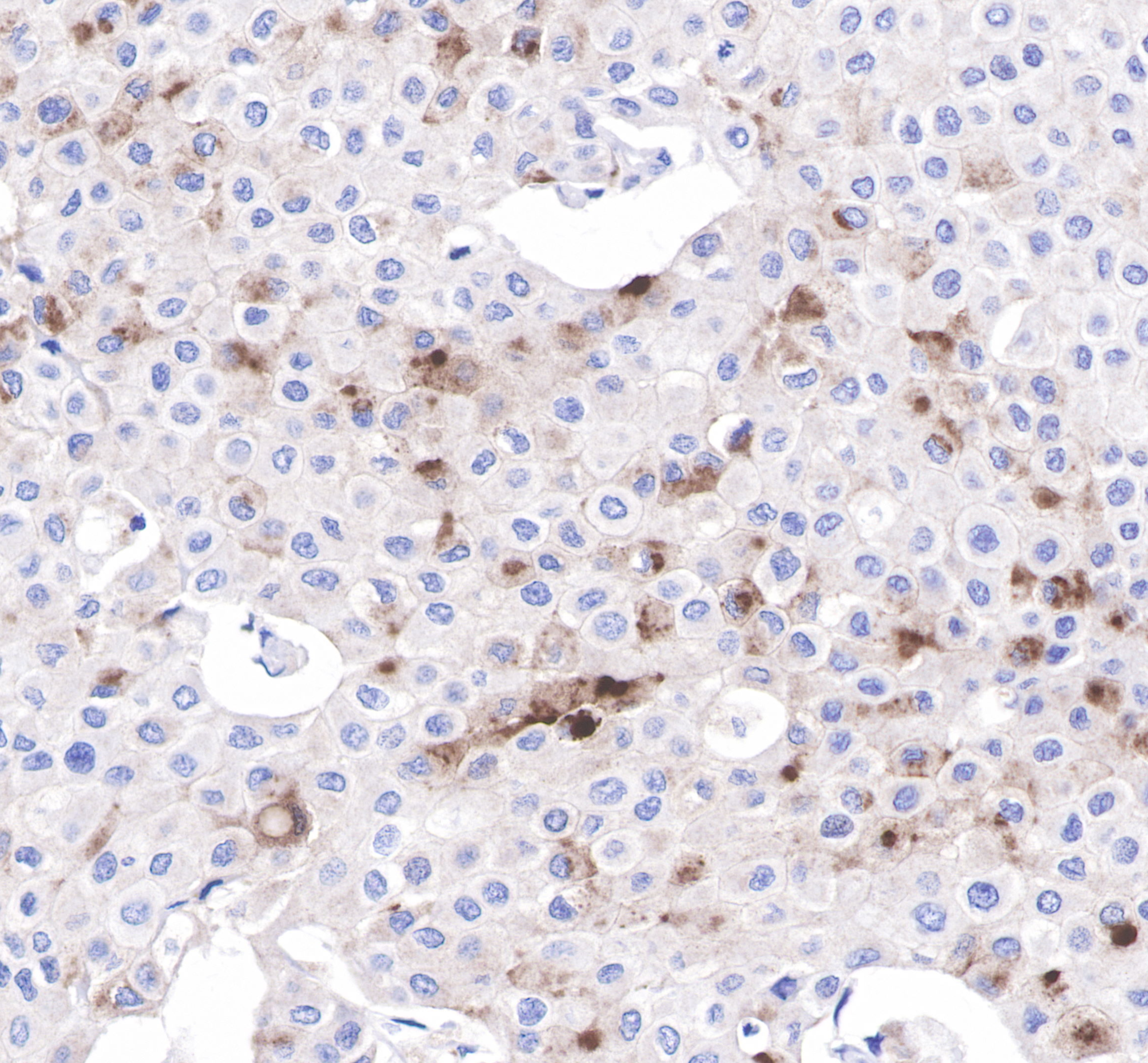
IHC shows cytoplasm and membrane staining in paraffin-embedded human liver. Anti-Keratin 8 antibody was used at 1/250 dilution, Secondary antibody: #abs20040. Counterstained with hematoxylin. Heat mediated antigen retrieval with Tris/EDTA buffer pH9.0 was performed before commencing with IHC staining protocol.
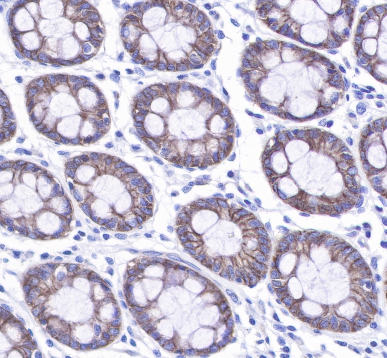
IHC shows cytoplasm and membrane staining in paraffin-embedded human colon. Anti-Keratin 8 antibody was used at 1/250 dilution, Secondary antibody: #abs20040. Counterstained with hematoxylin. Heat mediated antigen retrieval with Tris/EDTA buffer pH9.0 was performed before commencing with IHC staining protocol.
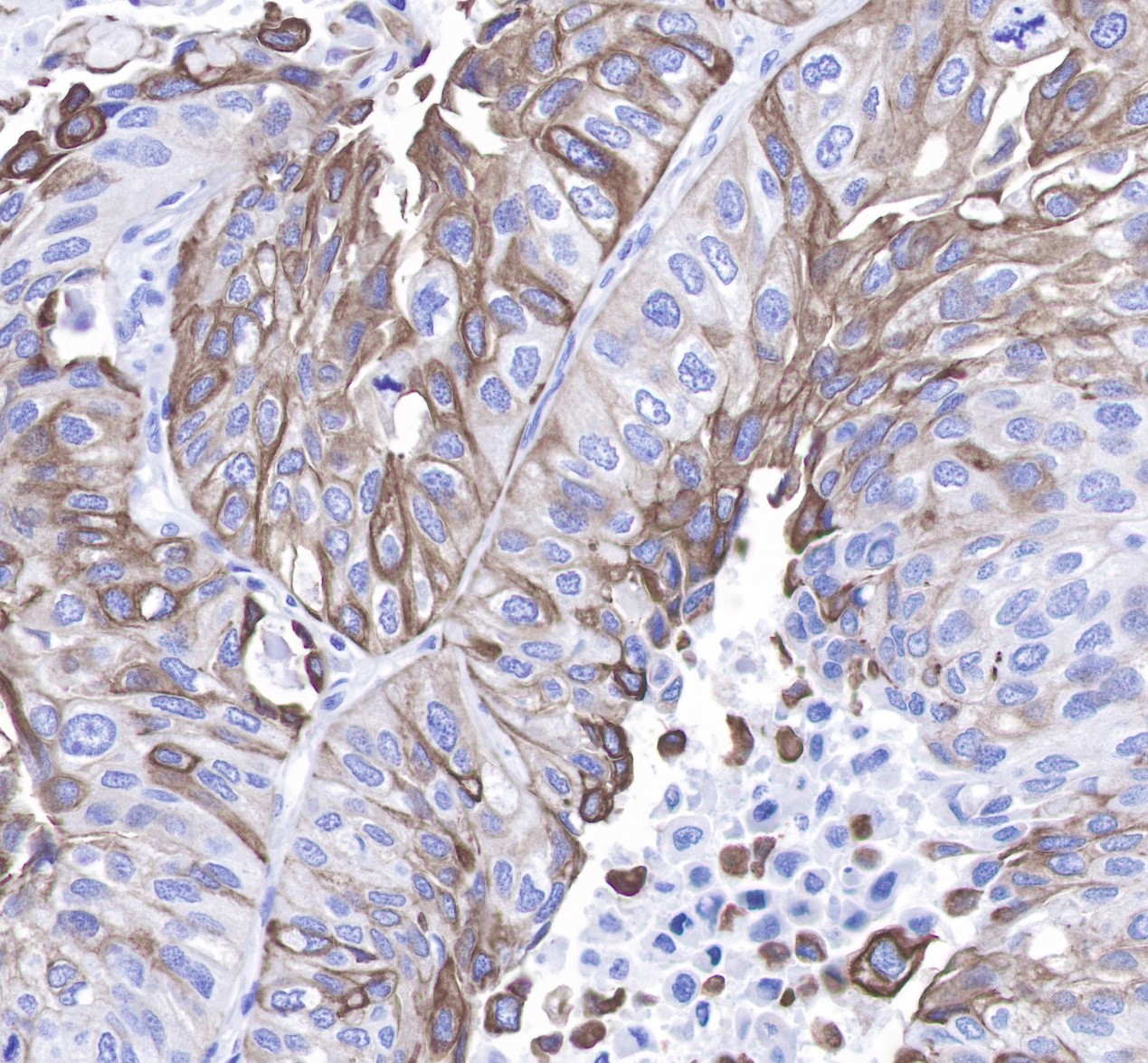
IHC shows cytoplasm and membrane staining in paraffin-embedded human lung squamous cell cancer. Anti-Keratin 8 antibody was used at 1/250 dilution, Secondary antibody: #abs20040. Counterstained with hematoxylin. Heat mediated antigen retrieval with Tris/EDTA buffer pH9.0 was performed before commencing with IHC staining protocol.
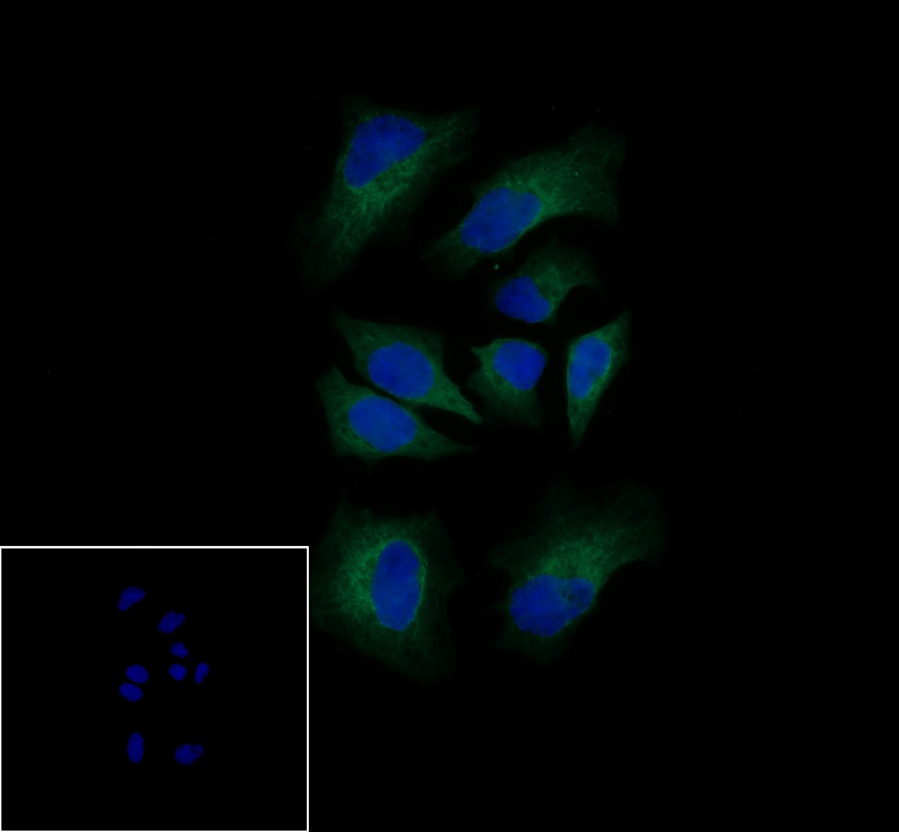
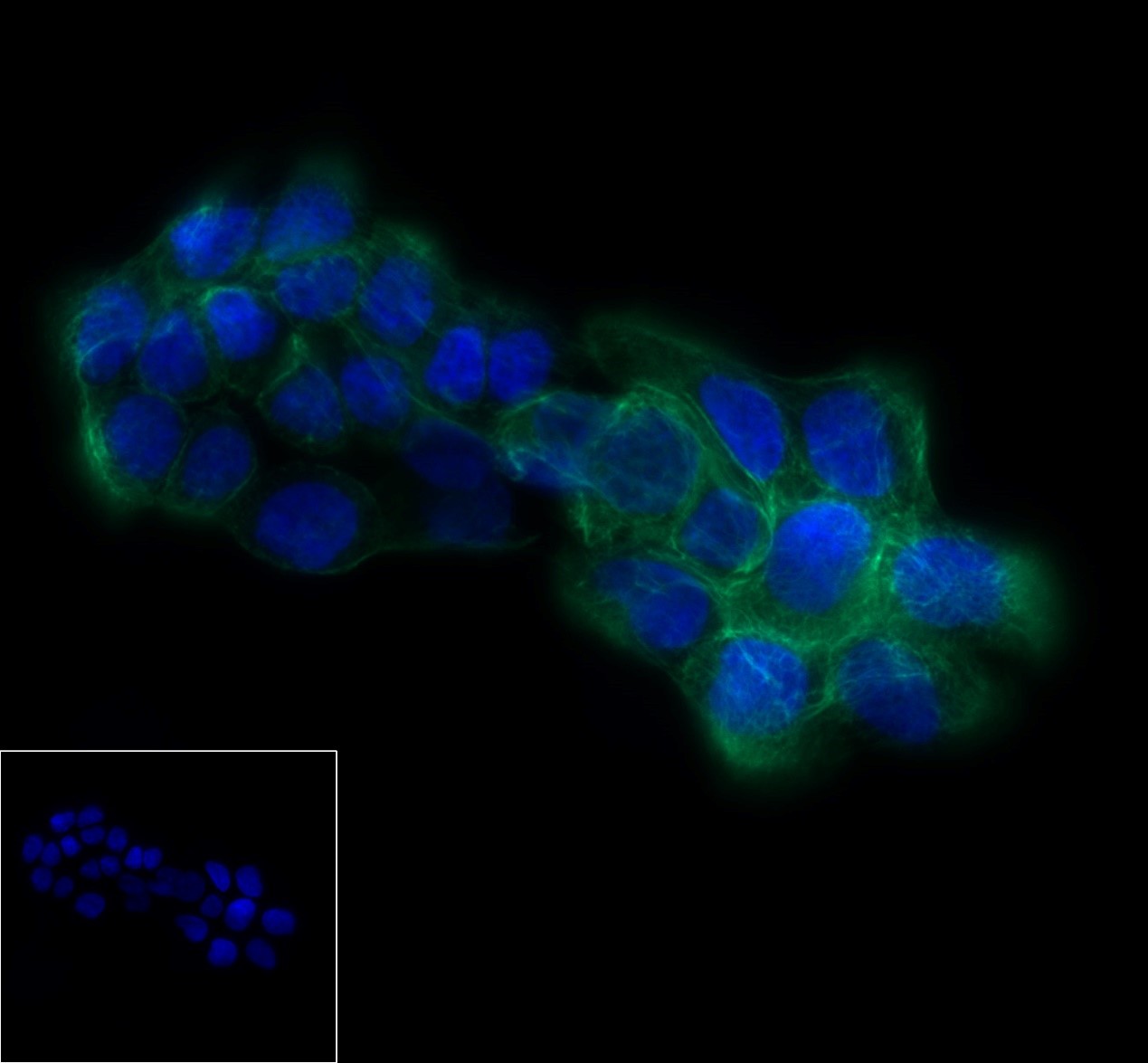
ICC shows membrane staining in HeLa cells. Anti-Keratin 8 antibody was used at 1/250 dilution and incubated overnight at 4°C. Goat polyclonal Antibody to Rabbit IgG - H&L (Alexa Fluor® 488) was used as secondary antibody at 1/1000 dilution. The cells were fixed with 100% methanol and permeabilized with 0.1% PBS-Triton X-100. Nuclei were countersained with DAPI.
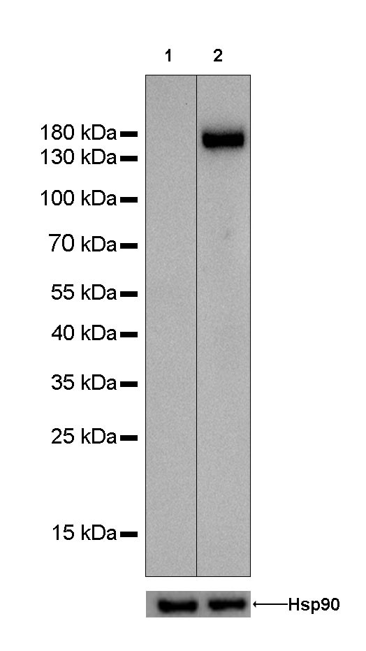
WB result of anti-CD31 antibody Primary antibody : Anti-CD31 antibody at 1/1000 dilution Lane 1 : Hela whole cell lysate 20 µg Lane 2 : THP-1 whole cell lysate 20 µg Negative control: Hela whole cell lysate Secondary antibody: #abs20040 at 1/10000 dilution Predicted MW: 82 kDa Observed MW: 150 kDa Exposure time: 103 seconds.
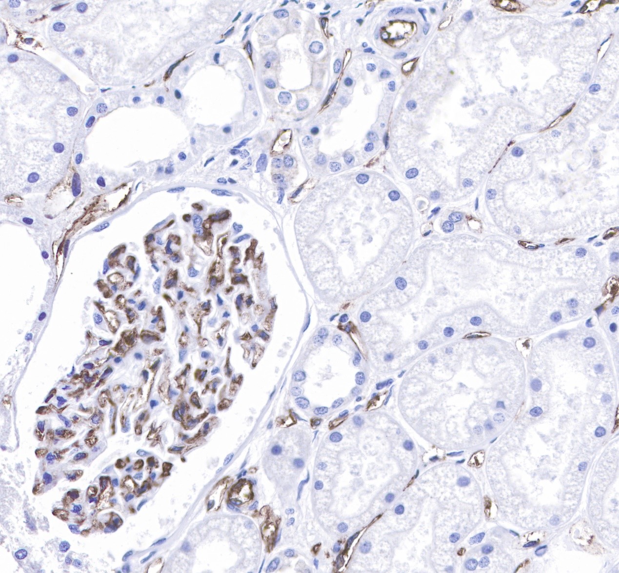
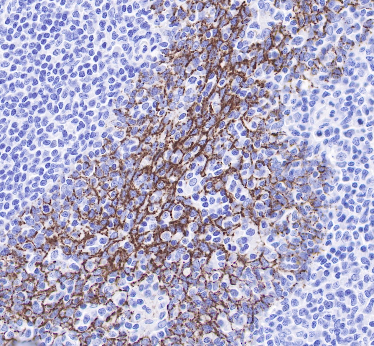
IHC shows membrane staining in paraffin-embedded human kidney endothelial cells. Anti-CD31 antibody was used at 1/250 dilution, Secondary antibody: #abs20040. Counterstained with hematoxylin. Heat mediated antigen retrieval with Tris/EDTA buffer pH9.0 was performed before commencing with IHC staining protocol.
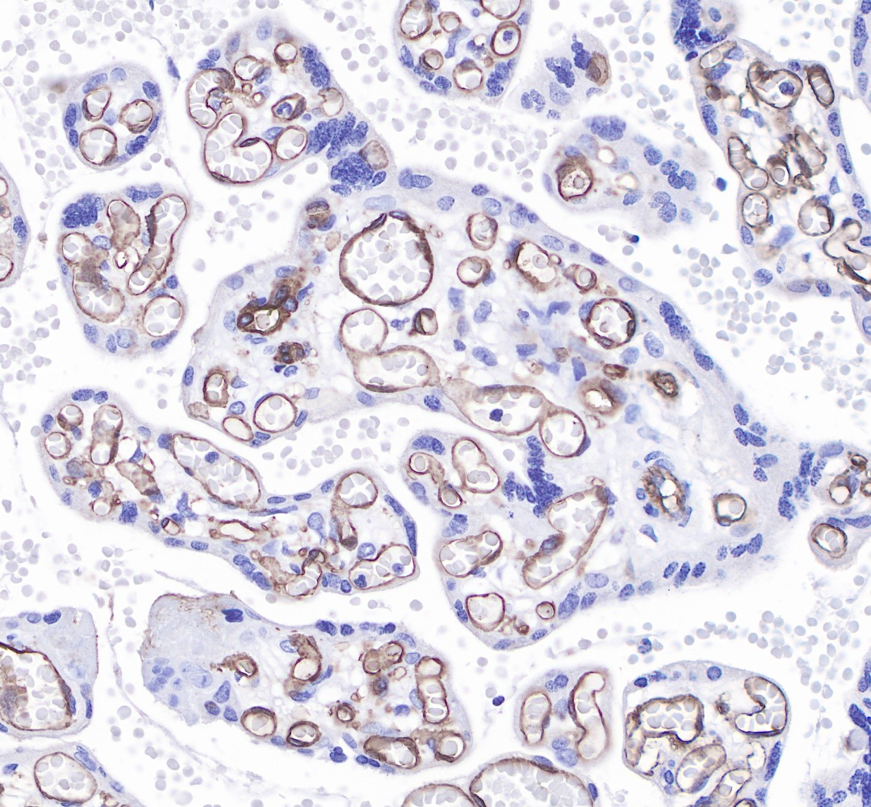
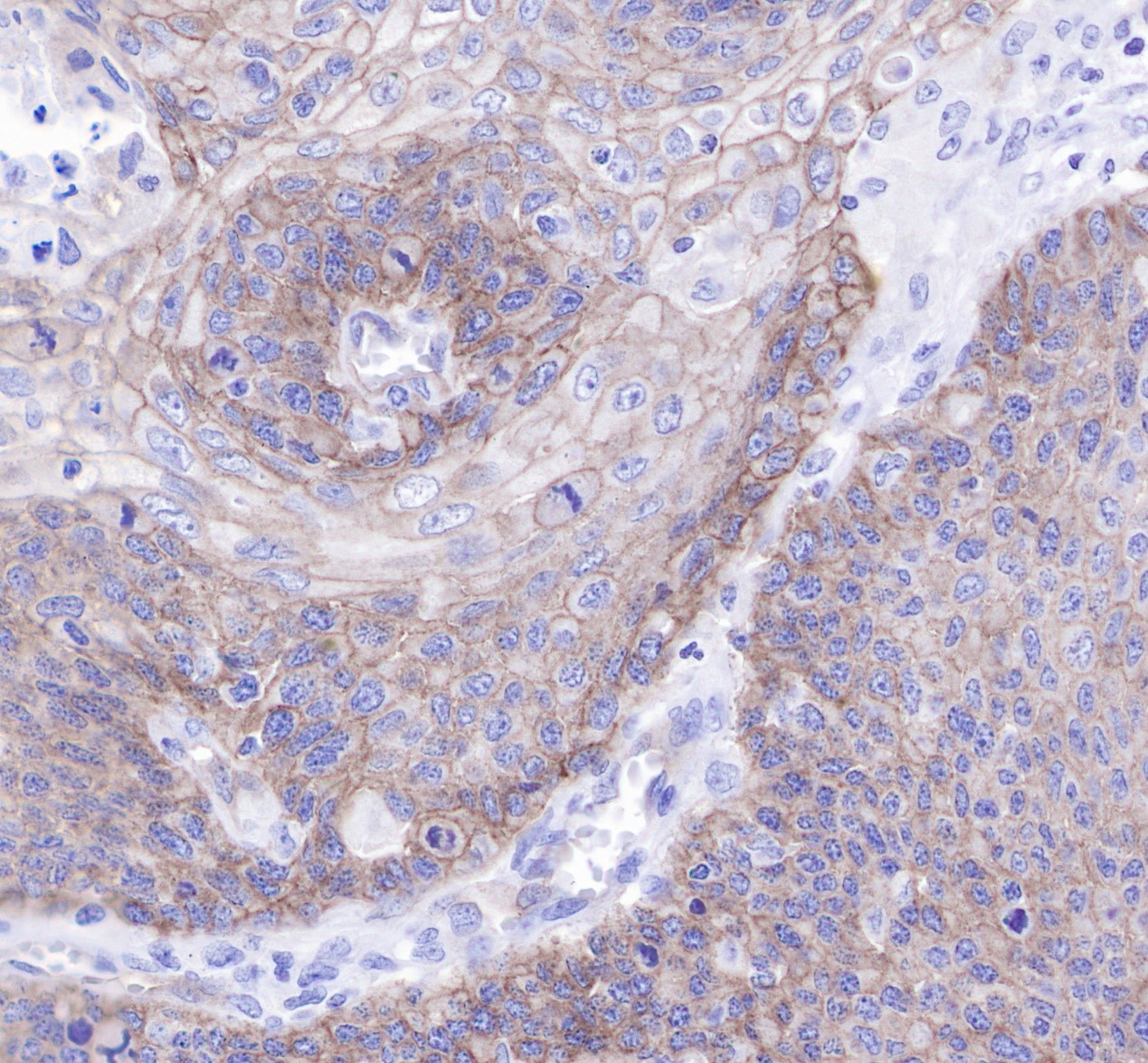
IHC shows membrane staining in paraffin-embedded human placenta endothelial cells. Anti-CD31 antibody was used at 1/250 dilution, Secondary antibody: #abs20040. Counterstained with hematoxylin. Heat mediated antigen retrieval with Tris/EDTA buffer pH9.0 was performed before commencing with IHC staining protocol.
Absin特色产品线:
WB相关:ECL发光液、预染marker、预制胶;IHC相关:二抗试剂盒、组化笔;IP/CoIP试剂盒;激动剂/抑制剂;血清、BSA、蛋白酶K、CTB、TTX、CEE;凋亡试剂盒;呼吸爆发试剂盒;ELISA试剂盒;重组蛋白;抗体: 二抗、标签抗体、对照抗体;定制服务(抗体/多肽/蛋白/标记/检测)...
爱必信(上海)生物科技有限公司
联系邮箱:info@absin.cn
公众平台:爱必信生物 .jpg)